Call Us
Goosnargh:
Lytham Road:
Kirkham:

IMPORTANCE OF DENTISTRY

IMPORTANCE OF DENTISTRY

At least once a year (as part of your horse’s annual health check), a thorough dental examination should be performed. This is particularly important for the winter months and for our older equine patients.

Dental disease can be very painful. Early detection and treatment of dental problems is vitally important in maintaining good oral health- we all know that prevention is better than cure! This will also enable is to advise with regards to feed types that will best suit your horse.

Common dental problems that we encounter when horses are struggling include:

  • Diastemata: these are gaps between the teeth where food can get trapped resulting in painful gum disease. This is the most common cause of quidding during the winter months.
  • Loose teeth: longer forage like hay requires more chewing therefore teeth which are slightly mobile can become problematic with these changes in feedstuffs.
  • Sharp points: these sharp points can cause ulceration of the gums and mucosa resulting in pain when eating or when ridden.
  • EOTRH (Equine Odontoclastic Tooth Resorption and Hypercementosis): this is a disease of the incisors resulting in severe pain. Horses can use their lips to graze during the summer months however struggle to pull hay out of haynets with these painful teeth.

If you have any concerns regarding your horse’s dental health or would like to arrange an examination with one of our vets, please give the practice a ring to book an appointment.

CASE STUDY: KISSING SPINES

CASE STUDY: KISSING SPINES

This 6-year-old WB mare first presented to vet Nikki in May, with a short history of bucking, napping and increasing resistance to going forward.

On examination, severe pain and spasm was noted over the back. No lameness was observed, but there was no transfer of energy from the hind limbs through the back, which was lacking in flexion. The horse tended to strike off incorrectly into canter or become disunited in this gait.

Gastroscopy revealed mild ulceration. Radiography revealed the presence of significant impingement of the dorsal spinous processes (colloquially known as ‘kissing spines’) of the vertebrae in the region under the seat of the saddle.

Given the potential for both gastric ulcers and kissing spines to cause the symptoms exhibited, the gastric ulcers were initially treated with Omeprazole and confirmed to be resolved one month later. This meant that treatment of the kissing spines could commence.

The X-ray findings in this horse can be present in clinically normal horses and in horses where kissing spines have been found to be the cause of pain, treatment can unfortunately fail to resolve an established pattern of evasive behaviour. Therefore, to be certain that this was the cause of the performance issues in this particular horse and that resolving the pain did indeed result in the horse performing normally, medical management was initially opted for.

This involved injecting the affected region of the back with steroid followed by a rehabilitative exercise programme and physiotherapy. Sometimes, medical management alone is enough to return horse to – and maintain them in comfort for their intended use.

Conversely, as was the case with this mare, the improvement is only short lived, and the performance issues return as the effects of the steroid start to wear off. ECVS surgeon Rosie therefore proceeded to surgically reduce the dorsal spinous processes, so they were no longer in contact. This procedure is known as a dorsal wedge ostectomy – amazingly, it can be carried out under standing sedation!

Once surgery was completed, the mare returned home to allow time for the surgical site to heal before an intensive non-ridden rehabilitative exercise programme was instigated (promoting correct muscling over the back).

Pre and post surgery

Ridden exercise was then carefully reintroduced (allowing the horse time to understand that ridden activity was no longer painful), and as you can see from the short video clips, the transformation from unhappy an unhappy, tense horse to a settled horse with powerful movement is remarkable. 

OAKHILL VETS CELEBRATES FIFTY YEARS

OAKHILL VETS CELEBRATES FIFTY YEARS

This year marks Oakhill Vets 50th anniversary, and we couldn’t be more excited to celebrate this momentous milestone!

It’s been quite a journey since 1974 – what began as a one-vet practice founded by Barry Johnson, now employs 130+ people over three divisions and three sites. We are proud to have achieved this remarkable development and growth, all whilst remaining fiercely independent.

On this special occasion, we take a moment to reflect on our accomplishments and most importantly the positive impact we have made to our patients and clients’ lives. We have consistently pushed boundaries and delivered exceptional care, while upholding our core values.

“Oakhill Vets remains dedicated to delivering the very best veterinary care to the local community,” said Tony Barry, CEO. “As we move forward into the future, we are excited for the next phase of developments which will enable us to continue to be at the forefront of animal care. We will continue to invest in the future, both in terms of technology and people.

“Our success is a testament to the dedication, hard work, and talent exhibited by our incredible team. It is due to their commitment and passion that we have reached this significant milestone. We recognise the positive impact we can make to the profession through supporting ongoing professional development and working with local universities and colleges, offering work experience to the next generation of vets.”

We have chosen StreetVet as our charity of the year and money raised from events throughout the year will go towards helping them to deliver free, essential veterinary care and service to people experiencing homelessness in the UK.

INVESTORS IN THE ENVIRONMENT AWARD: ACHIEVING ‘BRONZE’, AND HEADING FOR SILVER!

INVESTORS IN THE ENVIRONMENT AWARD: ACHIEVING ‘BRONZE’, AND HEADING FOR SILVER!

Last year, Oakhill Vets started our journey working towards ‘bronze’ accreditation with Investors in the Environment (iiE).

Investors in the Environment is a national sustainability accreditation that supports organisations to develop an ‘environmental management system’ that focuses on four key areas of sustainable development: Leadership and Governance, Climate Change, Nature and Natural Resources, and Pollution and Waste.

Being kind to the environment has always been a part of Oakhill’s ethos and working towards iiE accreditation has been a fantastic way to formalise our commitment to the environment. In working towards this accreditation, we wanted to challenge ourselves to make Oakhill’s operations as sympathetic as possible to people and the planet. It has given us the structure to hold ourselves accountable to reducing our carbon footprint and developing sustainable practices.

The accreditation has three levels – bronze, silver, and green. Achieving the bronze award is all about identifying resources that our company is going to measure and creating a base-line-year of data for these resources. As well as this, we needed to radicalise our environmental and sustainability policy, to include bolder aims, create a waste management plan, and produce a robust sustainability action plan, to set out a roadmap to achieving our sustainability goals.

Vet Lisa is the ‘Sustainability Lead’ at Oakhill, and she has been working hard, alongside the project’s sustainability champions, the wider staff team, and our directors, to complete all the necessary work to achieve this accreditation. After a busy period of reporting and planning, Oakhill had its ‘Sustainability Audit’ with the iiE team in October, and we’re very proud to announce that we achieved ‘bronze’ accreditation!

Next steps…

The next step is to begin working towards achieving ‘silver’ accreditation. This will build upon all the work we have done for the bronze accreditation and deepen our commitment to treating the planet with love, turning our sustainability goals into habits and practices. We are looking forward to the challenge!!

Case study: Mr T’s melanoma

CASE STUDY: MR T’S MELANOMA

Meet Mr T, who earlier this year developed a slight swelling on the left hand side of his back, under the saddle region. He didn’t seem overly bothered about this swelling, but his owner asked Sarah to check it out as they were concerned that it was steadily growing bigger.

Sarah was a bit perplexed – the swelling felt to be a solid lump under the skin attached to one of Mr T’s ribs. The lump was therefore ultrasound scanned, which showed it was a soft tissue mass and was spreading between the ribs and expanding down towards his chest cavity. Given its location and the fact that it was growing bigger and likely to be causing Mr T some discomfort, it was decided that it was best off being removed. A biopsy was taken to determine what the mass was and the degree of malignancy as this would determine the ‘margin’ required at surgery.

SURGERY

For his surgery, the team were assembled with Guy operating and Charlotte performing Mr T’s anaesthetic. Due to how close the mass was to his lungs, Mr T was fully anaesthetised, as any sudden movements on his part could have resulted in his thoracic cavity being inadvertently entered. With Mr T nicely asleep, Guy carefully dissected the mass away from the normal tissues surrounding it. A drain was placed to help reduce fluid build up where the mass had been removed, and once he was up and awake, Mr T had a belly bandage placed to help keep his surgery site clean. Thankfully, the mass was a melanoma, which is a relatively common tumour in older grey horses, albeit in Mr T it was in a very unusual spot!

MELANOMAS

Melanomas are typically found in the perianal region, under the tail, inside the sheath, around the vagina or inside the mouth, although some horses will develop them under the skin elsewhere or internally.

Melanomas themselves are not generally too much of an issue if located externally, unless they become very large and ulcerated and therefore sore, but removal is most straightforward whilst they are small. Also, there is some evidence that the fewer melanomas a horse has present, the less they will go on to develop, so early removal is now generally recommended.

Sycamore poisoning

SYCAMORE POISONING: THE FACTS

Many of you may have noticed the characteristic helicopter seeds present in sycamore trees as of late, so we thought it would be a good time to remind you about sycamore poisoning, also known as atypical myopathy.

WHAT IS IT? A highly fatal muscle disorder that occurs following the ingestion of sycamore seeds or leaves in autumn, or seedlings in spring, that contain the hypoglycin A toxin.

SEVERITY: There is sadly a 75% fatality rate with most non-survivors succumbing within 72 hours of the development of clinical signs.

PREVELANCE: The toxin is not present in all sycamores and toxin levels may differ at different times of year and under different climatic conditions. Cases often follow an adverse change in weather conditions such as frost or rain.

CLINICAL SIGNS: Weakness, stiffness, muscle tremors, a fast (sometimes irregular) heartbeat, difficulties in breathing and dark red/brown coloured urine. Other clinical signs include depression and signs of colic. Severely affected horses become recumbent and others may be found already deceased.

DIAGNOSIS

  • The presence of compatible clinical signs.
  • A history of grazing pasture containing sycamore trees.
  • Physical examination.
  • Blood work findings.

Blood work includes evidence of dehydration and an exponential increase in muscle enzyme values, with or without increased kidney enzyme values. We run this bloodwork in-house with a rapid turn-around time on results.

For the definitive diagnosis, sample(s) are submitted to the Royal Veterinary College (RVC) for analysis. Results take a number of days to return; therefore if atypical myopathy is suspected, then the affected patient will be treated as such.

TREATMENT

Time is of the essence and rapid initiation of treatment is essential to improve prognosis. Suspected cases are generally hospitalised to facilitate intensive medical management.

Treatment is predominantly based on supportive care including administration of large volumes of intravenous fluid therapy. The provision of adequate pain relief is vitally important and the administration of vitamins can be advantageous.

PREVENTION

Prevention is based on preventing exposure to sycamore seedlings in spring, and seeds and leaves in autumn. Fence off the sycamore trees and surrounding area.

  • Collect the seeds and leaves regularly.
  • Fields containing sycamore debris should not be used to make hay/haylage.
  • To discourage your horse from seeking out undesirable plants, additional forage should be provided if pasture is poor.

Important: helicopter seeds may travel up to 200 yards!

Remember, not all sycamores contain the hypoglycin A toxin. To determine if your trees do, you can get them tested at the RVC Comparative Neuromuscular Diseases Lab.

Canker

CANKER

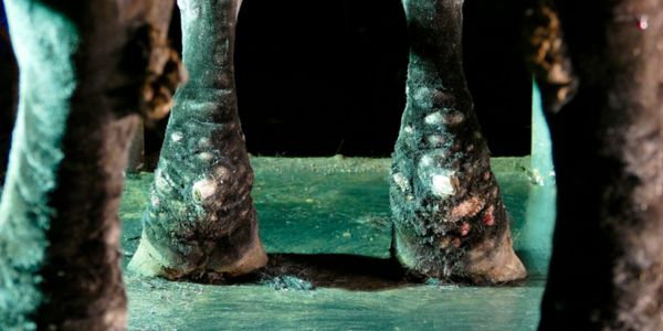
Max presented after his owner noticed the frogs of his feet had become soft and irregular. Upon examination it was clear that he was suffering from Canker in three out of four feet.

WHAT IS CANKER AND HOW DOES IT OCCUR?

Canker is an overgrowth of abnormal frog tissue, caused by bacterial infection, resulting in an over proliferation of soft, white sensitive frog and infectious material.

It is often described as being cauliflower-like with a cottage cheese discharge. It initially affects the back of the foot but can spread to affect the solar surface of the foot as well as deeper structures.

Fortunately, this condition is rare but when cases do occur it is usually in draught breeds like Max. The overgrowth of soft and sensitive material means the affected horses often become lame and treatment needs to be performed as soon as the condition is noticed for the best prognosis.

HOW IS CANKER TREATED?

Historically, corrosive agents have been applied to the lesions to burn away the abnormal tissue. However, this is unreliable and can cause a great degree of discomfort to the horse. Debridement (surgical removal) of the tissue back to normal appearing healthy frog is the best treatment method. For mild cases this can be done under standing sedation. However, in severe cases or if multiple feet are affected, debridement is performed under general anaesthetic, as in Max’s case.

Tourniquets were used on three of his legs to reduce the blood supply to the feet, thus reducing blood loss during surgery. Rosie and Stuart cut away all abnormal frog material until normal appearing healthy frog was reached, then his feet were bandaged to keep them clean.

Post surgery, Max’s feet were maintained in antibiotic dressings whilst normal frog tissue regrew and he was kept on pain relief to ensure he was comfortable throughout. After a couple of months, a few mini tidy ups of his feet under standing sedation, and tonnes of TLC he was finally given the go ahead to return to turn out and work.

Guttural pouch mycosis

GUTTURAL POUCH MYCOSIS

Ralph recently presented due to the development of mild head shaking behaviour and an abnormal respiratory noise when ridden. He subsequently developed an orange coloured nasal discharge but was otherwise well.

DIAGNOSIS

Endoscopic examination was used to determine the origin of the discharge. In Ralph’s case we could see that the discharge was exiting from one of his guttural pouch openings at the back of his throat. The image shows the fungal plaques covering the stylohyoid bone, cranial nerves and internal carotid artery.

Once inside the guttural pouch, a large volume of pus was noted in addition to white plaques which were adherent to the pouch lining. These findings were indicative of a fungal infection called guttural pouch mycosis.

We believe that Ralph’s headshaking and abnormal respiratory noise were secondary to the fungal plaques irritating the nerves within the guttural pouch.

Fortunately, guttural pouch mycosis is rare. Sadly, the disease can prove fatal in up to 50% of cases. The poor survival rate is due to fungal plaques breaking down the lining of the pouch, resulting in fatal bleeding or damage to nerves affecting the ability to swallow and breathe. It is not fully understood why or how this infection occurs and it can affect any horse of any age. Often the first presenting sign is a small nosebleed, which is why it’s important that we investigate nosebleeds in equine patients.

TREATMENT

Surgical treatment options are available that involve tying off the blood vessels to prevent severe bleeding. However, in Ralph’s case he was managed medically at our clinic. The team treated the affected pouch with an antifungal solution, in addition to removing the fungal plaques using forceps passed through the endoscope. He was given drugs to reduce inflammation and potassium iodide in his feed to increase the penetration of the antifungal wash.

Amazingly, on Ralph’s most recent assessment all of his fungal plaques had resolved. His signs of nerve irritation have largely improved, and the disease should not return.

Double trouble

DOUBLE TROUBLE

A couple of months ago Sarah attended the lovely Sally who had a surprise in store for all of us!

Sally was at stud to foal down and was noted to be going into the early stages of labour by stud owner Liz. However, it was soon apparent that she wasn’t progressing as expected – two legs had appeared but she was struggling and seemed exceptionally painful. Liz was quickly on the phone to vet Sarah who whizzed down to the stud as fast as Ribble Valley’s police find acceptable!

AN UNEXPECTED DISCOVERY

Upon feeling inside Sally, Sarah was able to identify one foal which was presented with their head and neck twisted over. However, it seemed very much stuck, despite being not overly large. A little more feeling further back revealed something no one was expecting – an extra pair of legs also within Sally’s pelvis – narrowing the gap and wedging the foal in place. At first Sarah wasn’t sure if these were the back legs of one very badly presented single foal, but after further palpation discovered that they were actually another set of front legs. It was at this point that the cavalry (Sarah’s second on call vet Julia) was called in to lend extra assistance if required.

SPRINGING INTO ACTION

Sally was quickly sedated, given pain relief, and an epidural placed in her spine to stop uterine contractions. Once contractions had stopped and she was more comfortable, Sarah was able to push the second foal further back beyond the pelvis, leaving room for the first foal to be repositioned and delivered. This was no mean feat and required both Liz and Sarah to pull on one leg each, as well as a lot of lubricant! Despite giving everyone a scare when she went limp half way through, Babette was soon wriggling round on the floor.

Julia arrived and was quite confused as to why Sarah was still two arms deep in Sally’s uterus, despite there being a foal on the floor, when Bettina (with her exit route now completely clear) made a swift entrance into the world. Due to being quite squashed up in the womb with her big sister, Bettina’s limbs were initially much weaker than hoped, and so Julia stomach tubed her with some colostrum in case she struggled to stand.

Babette and Bettina are now two months old and are both doing really well, full of character and sass. Sally has been a fantastic mother and is doing her best to look after both foals, ably assisted by stud owners Ian and Liz, who have worked tirelessly to supplement and support these twins.

TWIN PREGNANCIES

It is incredibly rare for a mare to carry twin foals to term; usually they resorb both pregnancies or will abort both foals at 8-9 months. It is even rarer for both to be delivered safely and for the mare to accept and be able to nurse both foals.

Normally if twin foals are seen when a mare is pregnancy scanned 14-17 days after covering, one of the embryos will be ‘squeezed’ in order to maximise the other foal’s (and the mare’s) chances of survival. We were incredibly lucky in this case that not only did Sally successfully carry both foals to term, but also that she was so closely monitored during her foaling, so that we were able to attend as soon as a problem was noted. If things had been left to chance, then the outcome would have been very different, with the possibility of losing not just the foals, but also the mare.

A calcinosis circumscripta conundrum

A CALCINOSIS CIRCUMSCRIPTA CONUNDRUM

Occasionally as a vet, there are cases that exercise our brains VERY hard! This was certainly the case with Zeus, a two year old gelding.

Zeus was found severely lame on his left hind limb whilst out grazing. On initial clinical examination, no localising signs could be found to explain the lameness. Intravenous pain relief was administered to facilitate returning Zeus to his stable. After 48 hours, his owners noted that some swelling had started to develop in the region of the stifle. Zeus was examined on yard to establish his fitness to travel, before coming into clinic for further investigations.

CLINIC INVESTIGATIONS

A large swelling within the muscle belly on the outer aspect of the limb was found 10cm below the stifle joint, which was severely inflamed.

X-rays revealed the swelling in the muscle belly to be a discrete mineralised mass, consistent with the appearance of a tumour, chronic abscess or calcinosis circumscripta. Radiography of the stifle joint itself was unremarkable.

Ultrasonography confirmed severe inflammation of the joint, with some damage to the stabilising ligamentous structures on the outer aspect of the joint. Ultrasonography of the mass revealed that it incorporated the overlying long digital extensor tendon.

Synoviocentesis (removal of synovial fluid) was performed on the joint. Instead of the small amount of transparent, straw-coloured fluid expected, an abundance of cloudy, cream fluid was obtained. This fluid underwent rapid analysis at Oakhill, where it was found to have a white blood cell count highly suggestive of infection of the joint although slightly bizarrely, no bacteria could be identified.

The question was, were the two findings (the muscle mass and the stifle joint inflammation) related? What was certain was that the extremely painful, inflamed joint required arthroscopic (keyhole) flushing under general anaesthetic to give Zeus a reasonable chance of recovery. Whilst the exact nature of the mass remained uncertain, it was decided that removal was the best plan of action.

WILL SURGERY PROVIDE MORE ANSWERS?

Equine surgery specialists Guy and Rosie operated together to minimise the length of the general anaesthetic. The mass was carefully excised, along with a portion of the affected tendon. The mass was found to contain a white, grit or chalk like substance – some of which appeared to be tracking up the tendon towards the joint! The joint was accessed separately via keyhole surgery allowing the abnormal fluid to be flushed out thoroughly and the thickened joint lining to be debrided. Much to the surprise of the surgeons, the same white chalky substance found in the mass appeared to be within the joint fluid as well. Zeus recovered well from surgery with the mass removed and a much healthier stifle joint.

The mass and synovial fluid were sent to an external lab for examination by a pathology specialist. This concluded that the mass was calcinosis circumscripta – an unusual condition characterised by the formation of a discrete, fibrous, mineralised mass under the skin, the cause of which is unknown.

Fluid analysis found white blood cell levels consistent with the initial findings at the clinic, but despite such high white blood cell levels normally only being recorded with joint infection, no bacteria were observed under microscope and no bacteria could be grown. The presence of calcified debris within the fluid was also confirmed.

Calcinosis circumscripta usually remains as a single defined mass. However, in Zeus’s case material from the mass appeared to have tracked up the tendon and penetrated the joint before causing extreme aggravation of the joint surface resulting in a severe inflammatory response.

Whilst Zeus remains a phenomenon of veterinary medicine, his recovery to date is extremely promising. He is now back enjoying being a youngster in the field with his friends prior to a check-up and medication of the joint at ten weeks post surgery!