Call Us
Goosnargh:
Lytham Road:
Kirkham:

Canker

CANKER

Max presented after his owner noticed the frogs of his feet had become soft and irregular. Upon examination it was clear that he was suffering from Canker in three out of four feet.

WHAT IS CANKER AND HOW DOES IT OCCUR?

Canker is an overgrowth of abnormal frog tissue, caused by bacterial infection, resulting in an over proliferation of soft, white sensitive frog and infectious material.

It is often described as being cauliflower-like with a cottage cheese discharge. It initially affects the back of the foot but can spread to affect the solar surface of the foot as well as deeper structures.

Fortunately, this condition is rare but when cases do occur it is usually in draught breeds like Max. The overgrowth of soft and sensitive material means the affected horses often become lame and treatment needs to be performed as soon as the condition is noticed for the best prognosis.

HOW IS CANKER TREATED?

Historically, corrosive agents have been applied to the lesions to burn away the abnormal tissue. However, this is unreliable and can cause a great degree of discomfort to the horse. Debridement (surgical removal) of the tissue back to normal appearing healthy frog is the best treatment method. For mild cases this can be done under standing sedation. However, in severe cases or if multiple feet are affected, debridement is performed under general anaesthetic, as in Max’s case.

Tourniquets were used on three of his legs to reduce the blood supply to the feet, thus reducing blood loss during surgery. Rosie and Stuart cut away all abnormal frog material until normal appearing healthy frog was reached, then his feet were bandaged to keep them clean.

Post surgery, Max’s feet were maintained in antibiotic dressings whilst normal frog tissue regrew and he was kept on pain relief to ensure he was comfortable throughout. After a couple of months, a few mini tidy ups of his feet under standing sedation, and tonnes of TLC he was finally given the go ahead to return to turn out and work.

Guttural pouch mycosis

GUTTURAL POUCH MYCOSIS

Ralph recently presented due to the development of mild head shaking behaviour and an abnormal respiratory noise when ridden. He subsequently developed an orange coloured nasal discharge but was otherwise well.

DIAGNOSIS

Endoscopic examination was used to determine the origin of the discharge. In Ralph’s case we could see that the discharge was exiting from one of his guttural pouch openings at the back of his throat. The image shows the fungal plaques covering the stylohyoid bone, cranial nerves and internal carotid artery.

Once inside the guttural pouch, a large volume of pus was noted in addition to white plaques which were adherent to the pouch lining. These findings were indicative of a fungal infection called guttural pouch mycosis.

We believe that Ralph’s headshaking and abnormal respiratory noise were secondary to the fungal plaques irritating the nerves within the guttural pouch.

Fortunately, guttural pouch mycosis is rare. Sadly, the disease can prove fatal in up to 50% of cases. The poor survival rate is due to fungal plaques breaking down the lining of the pouch, resulting in fatal bleeding or damage to nerves affecting the ability to swallow and breathe. It is not fully understood why or how this infection occurs and it can affect any horse of any age. Often the first presenting sign is a small nosebleed, which is why it’s important that we investigate nosebleeds in equine patients.

TREATMENT

Surgical treatment options are available that involve tying off the blood vessels to prevent severe bleeding. However, in Ralph’s case he was managed medically at our clinic. The team treated the affected pouch with an antifungal solution, in addition to removing the fungal plaques using forceps passed through the endoscope. He was given drugs to reduce inflammation and potassium iodide in his feed to increase the penetration of the antifungal wash.

Amazingly, on Ralph’s most recent assessment all of his fungal plaques had resolved. His signs of nerve irritation have largely improved, and the disease should not return.

Double trouble

DOUBLE TROUBLE

A couple of months ago Sarah attended the lovely Sally who had a surprise in store for all of us!

Sally was at stud to foal down and was noted to be going into the early stages of labour by stud owner Liz. However, it was soon apparent that she wasn’t progressing as expected – two legs had appeared but she was struggling and seemed exceptionally painful. Liz was quickly on the phone to vet Sarah who whizzed down to the stud as fast as Ribble Valley’s police find acceptable!

AN UNEXPECTED DISCOVERY

Upon feeling inside Sally, Sarah was able to identify one foal which was presented with their head and neck twisted over. However, it seemed very much stuck, despite being not overly large. A little more feeling further back revealed something no one was expecting – an extra pair of legs also within Sally’s pelvis – narrowing the gap and wedging the foal in place. At first Sarah wasn’t sure if these were the back legs of one very badly presented single foal, but after further palpation discovered that they were actually another set of front legs. It was at this point that the cavalry (Sarah’s second on call vet Julia) was called in to lend extra assistance if required.

SPRINGING INTO ACTION

Sally was quickly sedated, given pain relief, and an epidural placed in her spine to stop uterine contractions. Once contractions had stopped and she was more comfortable, Sarah was able to push the second foal further back beyond the pelvis, leaving room for the first foal to be repositioned and delivered. This was no mean feat and required both Liz and Sarah to pull on one leg each, as well as a lot of lubricant! Despite giving everyone a scare when she went limp half way through, Babette was soon wriggling round on the floor.

Julia arrived and was quite confused as to why Sarah was still two arms deep in Sally’s uterus, despite there being a foal on the floor, when Bettina (with her exit route now completely clear) made a swift entrance into the world. Due to being quite squashed up in the womb with her big sister, Bettina’s limbs were initially much weaker than hoped, and so Julia stomach tubed her with some colostrum in case she struggled to stand.

Babette and Bettina are now two months old and are both doing really well, full of character and sass. Sally has been a fantastic mother and is doing her best to look after both foals, ably assisted by stud owners Ian and Liz, who have worked tirelessly to supplement and support these twins.

TWIN PREGNANCIES

It is incredibly rare for a mare to carry twin foals to term; usually they resorb both pregnancies or will abort both foals at 8-9 months. It is even rarer for both to be delivered safely and for the mare to accept and be able to nurse both foals.

Normally if twin foals are seen when a mare is pregnancy scanned 14-17 days after covering, one of the embryos will be ‘squeezed’ in order to maximise the other foal’s (and the mare’s) chances of survival. We were incredibly lucky in this case that not only did Sally successfully carry both foals to term, but also that she was so closely monitored during her foaling, so that we were able to attend as soon as a problem was noted. If things had been left to chance, then the outcome would have been very different, with the possibility of losing not just the foals, but also the mare.

A calcinosis circumscripta conundrum

A CALCINOSIS CIRCUMSCRIPTA CONUNDRUM

Occasionally as a vet, there are cases that exercise our brains VERY hard! This was certainly the case with Zeus, a two year old gelding.

Zeus was found severely lame on his left hind limb whilst out grazing. On initial clinical examination, no localising signs could be found to explain the lameness. Intravenous pain relief was administered to facilitate returning Zeus to his stable. After 48 hours, his owners noted that some swelling had started to develop in the region of the stifle. Zeus was examined on yard to establish his fitness to travel, before coming into clinic for further investigations.

CLINIC INVESTIGATIONS

A large swelling within the muscle belly on the outer aspect of the limb was found 10cm below the stifle joint, which was severely inflamed.

X-rays revealed the swelling in the muscle belly to be a discrete mineralised mass, consistent with the appearance of a tumour, chronic abscess or calcinosis circumscripta. Radiography of the stifle joint itself was unremarkable.

Ultrasonography confirmed severe inflammation of the joint, with some damage to the stabilising ligamentous structures on the outer aspect of the joint. Ultrasonography of the mass revealed that it incorporated the overlying long digital extensor tendon.

Synoviocentesis (removal of synovial fluid) was performed on the joint. Instead of the small amount of transparent, straw-coloured fluid expected, an abundance of cloudy, cream fluid was obtained. This fluid underwent rapid analysis at Oakhill, where it was found to have a white blood cell count highly suggestive of infection of the joint although slightly bizarrely, no bacteria could be identified.

The question was, were the two findings (the muscle mass and the stifle joint inflammation) related? What was certain was that the extremely painful, inflamed joint required arthroscopic (keyhole) flushing under general anaesthetic to give Zeus a reasonable chance of recovery. Whilst the exact nature of the mass remained uncertain, it was decided that removal was the best plan of action.

WILL SURGERY PROVIDE MORE ANSWERS?

Equine surgery specialists Guy and Rosie operated together to minimise the length of the general anaesthetic. The mass was carefully excised, along with a portion of the affected tendon. The mass was found to contain a white, grit or chalk like substance – some of which appeared to be tracking up the tendon towards the joint! The joint was accessed separately via keyhole surgery allowing the abnormal fluid to be flushed out thoroughly and the thickened joint lining to be debrided. Much to the surprise of the surgeons, the same white chalky substance found in the mass appeared to be within the joint fluid as well. Zeus recovered well from surgery with the mass removed and a much healthier stifle joint.

The mass and synovial fluid were sent to an external lab for examination by a pathology specialist. This concluded that the mass was calcinosis circumscripta – an unusual condition characterised by the formation of a discrete, fibrous, mineralised mass under the skin, the cause of which is unknown.

Fluid analysis found white blood cell levels consistent with the initial findings at the clinic, but despite such high white blood cell levels normally only being recorded with joint infection, no bacteria were observed under microscope and no bacteria could be grown. The presence of calcified debris within the fluid was also confirmed.

Calcinosis circumscripta usually remains as a single defined mass. However, in Zeus’s case material from the mass appeared to have tracked up the tendon and penetrated the joint before causing extreme aggravation of the joint surface resulting in a severe inflammatory response.

Whilst Zeus remains a phenomenon of veterinary medicine, his recovery to date is extremely promising. He is now back enjoying being a youngster in the field with his friends prior to a check-up and medication of the joint at ten weeks post surgery!

Iris cysts

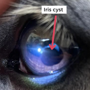
Iris cysts are a common finding in the equine eye. More often than not, they do not cause any issues and are an incidental finding. Some; however, can grow to a size where they begin to affect a horse’s vision. Such horses generally present to us due to spooky behaviour. This is most apparent in sunny conditions, when the horse’s pupil is small and the cyst blocks light rays from reaching the back of the eye.

Iris cyst

Treatment options

We will recommend treating iris cysts if we feel they are clinically significant – the horse in question is showing visual deficits or has started to spook.

Traditionally the treatment for iris cysts was laser ablation. We recently undertook a new technique to drain an iris cyst called ‘standing trans-corneal aspiration.’ This technique, which can be performed in suitable candidates, is performed under standing sedation, and local anaesthesia of the eye. A very thin needle is inserted into the eye, and the fluid from inside the cyst drained. The patient requires minimal medication following the procedure and most tolerate it really well.

If you would like to know more about iris cysts, please get in touch to discuss it with one of our equine vets.

Foaling

Breeding season is in full swing and the team have been busy seeing lots of beautiful mares and foals.

When it comes to the actual birth, the biggest difference between horses and other species, such as cattle, is the speed of foaling. Once stage 2 (active labour) has commenced, the foal should be born within 15-45 minutes. Foals ‘dive’ out of the mare with two front feet followed by the muzzle. You should call your vet within 10 minutes of active contractions beginning if the foaling isn’t progressing, or any issues are noted. There are a variety of options available to deliver the foal, from manually repositioning to a surgical caesarean section (which fortunately is incredibly uncommon).

Staging of foaling

Stage 1 – 1-4hrs – The mare is preparing for foaling, and starts to become restless, pawing, pacing and getting up and down. She may sweat up and frequently urinate and defecate.
Stage 2 – 15-45 minutes – This is active labour, when the mare’s waters break, contractions begin and the foal is delivered.
Stage 3
– 2-4 hrs following birth – Passing of the placenta

Two complications we see during foalings are red bag deliveries and retained foetal membranes (retained placenta). These are two conditions at the opposite ends of the spectrum involving the release of the placenta.

Red bag delivery

In a red bag delivery mares will deliver the foal within the placenta, rather than it bursting to release the foal. A red velvety bag is seen first, before the foal or any fluids. THIS IS AN EMERGENCY SITUATION as once the placenta has separated from the mare, the foal will not be receiving any oxygen. The foal requires immediate delivery. The ‘red’ bag needs to be broken open using your fingers or scissors. If both the foal’s feet and nostrils are present we recommend pulling to help the foal out quickly to give it the best chance of survival.

Retained placenta

A placenta is classed as ‘retained’ when it is still attached to the uterus (womb) 3–4 hours post-partum. At this point, veterinary advice should be sought as horses are very sensitive to toxins released from bacteria on a retained placenta. These toxins can then cause endotoxemia, colic and laminitis. As vets we usually give these mares a combination of antibiotics and anti-inflammatories, to reduce the risk of endotoxemia. We also administer drugs to stimulate the uterus to contract, flush the uterus, and finally apply gentle traction on the placenta. In some circumstances it can take several visits, and over 24 hours before the placenta has loosened sufficiently to be removed. The mare may require antibiotics and uterine flushing for several days thereafter.

Foal targets

  • 1 hour – Standing
  • 2 hours – Nursing
  • 3 hours – Passed meconium (first droppings)
  • 24 hours – Antibody (IgG) blood sample

Once the foal has been born, we expect them to stand and nurse within a couple of hours. This is particularly important as foals are born without antibodies which protect them from diseases in the environment. The mare’s colostrum contains antibodies, but the foal quickly loses the ability to absorb them. Maximum absorption occurs during the first 12 hours of life, with the foal’s gastrointestinal tract gradually losing the ability to absorb antibodies such that by 24 hours absorption has ceased.

We can check the foal has received sufficient antibodies with a blood test taken at the mare and foal’s 24-hour post-partum check. If the antibody levels are insufficient, we can administer hyperimmune plasma to boost the antibody levels to a protective level. This will protect the foal from life threatening conditions such as sepsis.

Planning on breeding your mare this year? Call us on 01772 861300 to request a FREE copy of our Mare & Foal Guide or to discuss the options available with one of our experienced vets.

Has your horse got itchy skin?

As the weather improves and pollen levels, as well as insect numbers increase, allergic skin disease is seen more frequently. Horses with allergic skin disease can present with itchiness or hives or both itchiness and hives together.

Sometimes it is easy to identify the trigger but more often than not, the cause is not easy to identify, and management of these horses can be incredibly frustrating with flare-ups common.

Management amendments can be made and the response to these changes monitored but in recurrent cases that we are struggling to control, skin tests (IDAT- intra-dermal allergen testing) can be performed to identify allergens which may be contributing to allergic skin disease. Skin testing remains the ‘gold standard’ diagnostic test for horses with allergic skin disease at this time.

Intra-dermal allergen testing

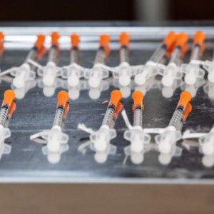
We are fortunate to be able to offer this service to our patients at Oakhill. Small blebs of common allergens are injected intra-dermally and the response to the allergens is checked 30 minutes, 4 hours and 24 hours following injection.

IDAT

We subsequently provide our clients with a full written report detailing changes in management which may assist in managing these patients based on the results of the individual patient’s skin tests. De-sensitising vaccinations can also be formulated based on results of the skin tests to further assist in the management of these cases.

If you have any questions regarding allergic skin disease or the diagnosis and management of, please do not hesitate to contact the team.

Dental Case Report

Isabel presented to Stuart after her owners noticed that she had separated from her field mates. Once brought into the stable she was quidding and unable to eat haylage.

X-ray of horse Isabel's teeth

On examination, a significant swelling of Isabel’s right lower jaw was noted. As we were suspicious of a dental issue, after antibiotics and anti-inflammatories were administered to control her temperature and provide some comfort, radiographs (X-rays) were taken. This revealed a tooth root infection of the 3rd cheek tooth on her right lower jaw. The infection had caused lots of bony reaction with thinning and loss of parts of the jaw – the red arrow on the radiograph points towards this.

Once Isabel was stabilised and at the clinic, we proceeded with extracting the infected tooth under standing sedation and local anaesthesia (a nerve block). The tooth came out in one piece and as you can see, it was a whopper!!

Tooth that had been extracted from horse Isabel

Tooth root infections commonly occur in youngsters just several months after tooth eruption. Although Isabel now has one less tooth in her mouth, she is pain free and was very quickly back to eating happily as you can see.

Isabel the horse

Respiratory tract health

Does your horse cough at the beginning of an exercise session? Do you often find accumulations of mucus outside your horse’s stable door? These subtle signs can indicate lung inflammation.

Formerly known as COPD or RAO, Equine Asthma is commonly seen in equine practice. For the purpose of simplicity, two forms are recognised- Summer asthma and the more traditional dust-induced form.

Both forms of the disease occur when a susceptible horse inhales either an allergen, to which they are allergic, or dust into their airway. This results in the airway spasming and the production of increased amounts of both inflammatory cells and mucus within the airway.

Affected horses present with clinical signs of varying degrees of severity. Some horses present with the subtle signs mentioned above or with a history of poor performance, whereas others present in respiratory distress and struggling to breathe.

On examination affected horses will generally have an increased breathing rate and effort combined with nostril flare. Mucoid nasal discharge may also be present. More severely affected horses may cough, have abdominal effort to their breathing and may have a ‘heave-line.’ A horse’s history combined with clinical examination findings will point towards a diagnosis of asthma but for definitive confirmation, airway endoscopy, to visualise the airway and grade airway mucus combined with laboratory analysis of respiratory tract samples is required. This will also rule in/out secondary bacterial infection.

Treatment of asthma should be based on environmental modifications plus drug therapy on an as needed basis. Horses affected by dust-induced asthma should be turned out in so far as possible. The stable environment should be closely examined. Ideally, the stable will have an inlet and outlet for airflow. Forage should not be stored adjacent to the stable to minimise the dust to which the patient is exposed. Horses should be groomed outside of the stable and fresh bedding laid when your horse is not in the stable environment. If your horse’s stable is unsuitable, try find an alternate stable on your yard that has better airflow. If feeding hay or ‘dry’ haylage, steaming is optimal to reduce dust particles but if not available, soaking should be considered. Managing Summer asthma is more challenging, but the measures outlined above should be followed to ensure stable ‘air hygiene’ is a good as possible.

Drug therapy, when needed, is based on relieving airway spasm (bronchodilators) and reducing airway inflammation and mucus production (steroids and mucolytics). Drug therapy can be provided by the oral or inhalatory routes. Oral medication includes bronchodilators, steroids and mucolytics (which serve to break-up airway mucus). From an inhalatory medication perspective, three options are available- the Equihaler, MDIs and nebulisation. The Equihaler is a licensed, steroid based product which utilises a fine mist to deliver steroid directly to the lungs. The product is very safe as the steroid acts at the lung surface only. We have had some great success using this product in cases of severe asthma and in those that did not respond to other forms of medication. MDIs, human asthma inhalers administered via a baby asthma face mask, have been utilised in equine practice for many years. These are probably most useful in relatively mild cases. Drug delivery via a nebuliser is the third inhalatory option but arguably used less frequently these days.

If you have any questions regarding asthma or think your horse may be affected, please do not hesitate to contact the team for guidance.

What is MRI used for?

Our standing MRI scanner can be used to assess injuries from the hoof, up to and including the hock and carpus (knee). The scanner is specifically designed to image the lower limb in the standing horse, as this is the most common site of lameness.  It has revolutionized our understanding of the structures of the hoof, and we can now differentiate between the multiple conditions that were encompassed as ‘navicular syndrome’.

Previously, a horse with forelimb lameness that was localized to the foot, was diagnosed with ‘navicular syndrome’.  However, a lot of the time, the severity of the lameness did not fit with the mild observations noted on radiographs (X-rays). We now know, through the use of MRI, that there are many other anatomical structures that could be injured and causing the lameness. With forelimb lameness being a common problem in horses, this diagnostic imaging tool means we can target rehabilitation, farriery, and treatment more specifically.

Injuries identifiable on MRI would include deep digital flexor tendon lesions within the hoof. Without the use of MRI this condition would have been misdiagnosed, leading to inaccurate management and unsoundness. MRI can also assess ligaments within the hoof capsule, such as the collateral ligaments of the coffin joint, which are often painful when horses are lunged in a circle. This amazing imaging modality also shows us the degree of inflammation within synovial structures such as the coffin joint and navicular bursa of the foot which cannot be visualized in any other way. Not only does MRI allow us to diagnose more accurately, but it allows us to monitor the progression of conditions and carefully assess the horse’s response to treatments.

X-ray imaging is used to assess bone pathology as an initial tool. However, it can take up to 2 weeks following injury before the bone pathology is noticeable on radiographs, and sometimes it is not visualized at all.  MRI is the only imaging modality that can assess inflammation within bones such as bone bruising or cysts. These can cause severe lameness and require long periods of rest but would not be diagnosed without the use of MRI.

As equine vets we are eternally grateful for these advancements in technology which have enabled us to achieve an accurate diagnosis much faster than ever before, and as we know, a faster diagnosis leads to more precise treatment and management protocols to get your horse feeling in tip-top shape again.