Call Us
Goosnargh:
Lytham Road:
Kirkham:

Grass Sickness in horses

Grass Sickness is a disease of horses, ponies and donkeys in which there is damage to parts of the nervous system which control involuntary functions, producing the main symptom of gut paralysis.

Also known as Equine Grass Sickness (EGS) the cause is unknown but the nature of the damage to the nervous system suggests that a type of toxin is involved – potentially botulism neurotoxin acquired from soil.

The toxin may also affect nerves supplying other body systems resulting in other signs of EGS such as droopy eyelids, inability to swallow & muscle tremors to name but a few.

Three forms of the disease have been reported: the acute, subacute and chronic forms. The form a patient succumbs to depends on the extent of nerve damage.

Horses affected by the acute form of the disease present showing signs of colic often indistinguishable from other forms of colic meaning that it may be suspected that the patient has a twisted gut or other form of surgical colic.

As a result, such patients often undergo colic surgery and the diagnosis of EGS is often made presumptively on the surgery table. This form of the disease is 100% fatal.

In horses with subacute or chronic EGS, the time course of the disease is more gradual and these patients may present with a high heart rate, mild episodes of colic, a tucked up appearance, an inability to swallow, drooling saliva, droopy eyelids, muscle tremors and patchy sweating. This form of the disease is also fatal.

Horses with the chronic EGS may survive but require intensive management to maintain hydration and nutritional requirements. The likelihood of survival depends on the extent of nerve damage.

The only way to definitively diagnose EGS is to examine an intestinal biopsy. Surgery is required to obtain a biopsy. Therefore, horses are frequently diagnosed based on the presence of compatible clinical signs.

Vets often perform an eye drop test known as a phenylephrine test. When these drops are applied to one of the patient’s eyes the droopy eyelid appearance improves. Other conditions may also cause droopy eyelids so this test is by no means perfect.

Horses at risk of succumbing to EGS include any horse at grass but the condition is most commonly seen in young animals aged between 2 & 7 years.

Cases have been reported throughout the year but occur most frequently in late spring/early summer. Overweight horses are also at increased risk. Other reported risk factors include recent soil disturbances, overuse of ivermectin based wormers, a recent change in pasture & being at grass 24/7.

Prevention is based on avoiding changes in management, especially in youngstock, at the ‘at risk’ time of year. Soil disturbances should also be kept to a minimum. Ideally, horses should be stabled for at least part of the day and offered hay or haylage. Furthermore, overuse of ivermectin based wormers should be avoided and ideally, a wormer containing an alternative drug should be used prior to turn out. Co-grazing with sheep or cattle may also be protective.

When a case has been diagnosed at a property,it is of paramount importance to stay calm and to avoid any sudden changes in management. In our opinion, in-contact horses should not be moved field as moving pasture is itself a risk factor for

EGS. Furthermore, fields within a 10km radius are theoretically ‘at risk.’ Co-grazing with a patient that has succumbed to EGS may itself be protective suggesting an acquired immunity. We would; however, recommend that young horses are kept off an affected field during future grazing seasons.

A vaccine trial is currently underway which, if licensed, will hopefully provide us with an effective means of preventing EGS in the future.
Immunity in foals

At birth, foals are immuno-naive meaning that they are born without any protective antibodies (immunoglobulins/IgG) against common ‘bugs’ in their environment.

In order to gain protection, foals need to ingest a sufficient quantity of good quality colostrum (first milk containing high levels of antibodies) within the first 12 hours of life.

It is during this time that foals have maximal absorption of ingested IgG. After this time, absorption rapidly decreases.

Adequate IgG levels are crucial to protect foals from developing infections (namely sepsis). Inadequate levels result from failure to ingest sufficient quantities of colostrum or poor quality colostrum (e.g. when the mare runs milk before birth and loses IgG).

To determine that foals have adequate IgG levels, we recommend measuring IgG using a simple blood test, generally 18-24 hours following birth.

If levels are inadequate then a hyperimmune plasma transfusion is recommended to provide the foal with IgG and in turn protect the foal from developing life threatening infections.


Image source: foalpatrol.com


Tooth decay in horses

Similar to humans, horses’ teeth suffer from decay, referred to as ‘caries’.

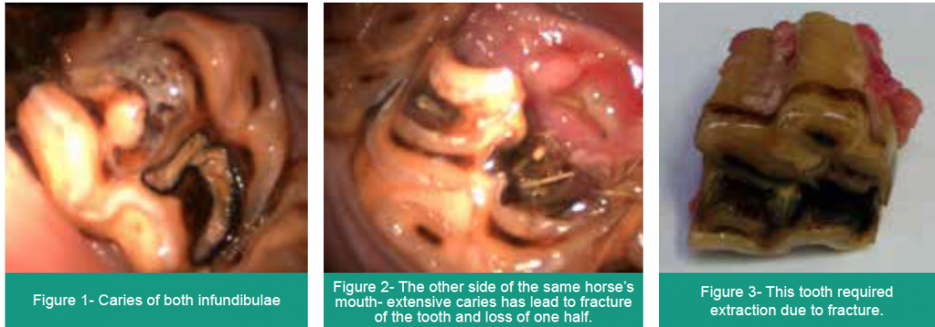
Horses’ upper cheek teeth have infundibulae, cup-shaped invaginations in their grinding surface that are normally filled with a hard material called cementum. Sometimes these infundibulae are incompletely filled during development, leaving a cavity for food to settle in and decay. Over time the decay weakens the structure of the tooth, predisposing it to fracture and, if the decay advances beyond the margins of the infundibulum and into the sensitive pulp system, an apical (root) infection may arise. Both of these situations require extraction of the tooth.

Caries of the infundibulae

How can we prevent this?

Infundibular caries cannot be detected by feel, therefore is it imperative that every horse’s mouth is visually assessed using a headtorch during routine dental examinations to detect these before advanced disease develops.

It is possible to perform cleaning and filling of the cavity with filling material. This is termed an ‘infundibular restoration’. We now have the equipment to perform this at our clinic.

To discuss any of our Advanced Dentistry services, please call Sam Baker at the surgery: 01772 861300

Thinking about breeding your mare?

Pre-Breeding Testing

Taking your mare to stud? Depending on which stud you are going to, you may be asked to test for a variety of diseases.

The most common disease is contagious equine metritis (CEM). This is a bacterial uterine infection caused by T. equigenitalis, K. pneumoniae or P. aeruginosa. Testing involves takes a swab – either clitoral or endometrial (if the mare is in season) – which is then cultured (grown) for at least 7 days, or tested by PCR which gives a result in 1-2 days. The PCR option is slightly more expensive.  

Next is EVA or Equine Viral Arteritis. This disease can cause a variety of signs including fever, lethargy, and swelling of eyes, limbs, scrotum and mammary glands. In rare cases it can be fatal. It is spread by both the respiratory (droplets in the air) and venereal (mating) routes. If stallions are infected, they can become chronically infected for years, spreading it to many mares via infected semen. Testing is performed by blood sampling.

Then, there is also Equine Infectious Anaemia (EIA). Horses with EIA are often extremely sick, as the disease damages their red blood cells causing an anaemia. It is spread by the transfer of infected blood, colostrum and semen. Again, this is checked for via a blood sample.

CEM, EVA and EIA are all notifiable disease. The last reported case of CEM in the UK was in February 2022, while the last reported EVA was 2019 and EIA was 2012.

Finally, it is worth mentioning strangles. Whilst this isn’t necessarily related to breeding outbreaks can occur when horses from different sources meet at stud farms. Therefore, some studs require testing, in the form of a blood test, prior to arrival at stud.

As different studs have different test requirements, we recommend that you check your stud’s requirements prior to your veterinary appointment for testing.

Twins

Twins are a problem in horses. This is because only in exceptional cases can a mare give birth to two healthy foals. Should a twin pregnancy continue, sadly most mares will lose both pregnancies mid-late gestation due to lack of space and nutrition for both foetuses.

We can minimise the risk of this by ultrasound scanning mares after covering/insemination. We would usually scan mares between 14-20 days to look for twins. At this stage it is easier to identify them and manage them. If detected, we will try to abort one of the embryos then recheck a week later to see if the remaining embryo is still healthy.

Artificial Insemination Options

Fresh semen – This is used within 3 hours of being collected. It is the most successful option, as the semen will live for up to 3 days in the mare, meaning the timing of insemination is less critical. However, it is limited to local stallions reducing the options available.

Chilled semen – This needs to be inseminated within 48hrs. It is usually collected and then posted overnight to the mare in a special container. This limits it to stallions within the UK and Europe.

Frozen Semen – After collection, the semen is frozen in liquid nitrogen at -196°C. It can be stored for years after collection, and hence can be shipped around the world. It also means the stallion does not need to be at stud when you’re breeding. However, fertility rates are lower, and the semen only survives in the mare for 6hrs so the timing of insemination with ovulation is crucial.

Along with different semen options, there are some considerations as to its place of origin.

UK semen – With UK semen, there tends to be less postage delays and no issues with regards to customs. If semen is required urgently, then it can be collected in person.

Foreign semen – With semen originating abroad, there is a higher risk of delivery delays meaning missed cycles. Since Brexit there has been an increase in problems with health paperwork and customs delays. However, semen from a much larger range of stallions is available giving you more choice regarding how to produce your perfect foal!

Sweet Itch

Sweet itch is a common disease of horses in summer causing them to rub their mane, tail and body. It is caused by a hypersensitive reaction to the saliva of the female Culicoides midge. Affected patients can suffer from frenzied itching of the mane, tail, head, poll and abdominal areas. This results in loss of hair/fur, skin thickening and in severe cases, open wounds from self-trauma.

How can you manage it?

There are three main approaches to approaching/managing sweet itch: midge avoidance, soothing creams/shampoos and medications.

Avoidance is better than cure so the most effective method is midge avoidance. This includes:

  • Stabling mid-afternoon to mid-morning, as midges are most active dawn to dusk.
  • Using fans in the stables to help reduce midges.
  • Choosing open (windy) fields, avoiding woodlands and areas of standing/stagnant water.
  • Keep muck heaps away from grazing.
  • Using full fly rugs, to provide a physical barrier.
  • Fly/midge repellents, especially those with contain permethrin or cypermethrin which should last a couple of days- we can provide small bottles of these on request, which will dilute to 500ml of repellent.

In mild clinical cases, topical management through the use of soothing creams/shampoos on affected areas is generally the first port of call. While these products do not treat the problem, they improve the comfort of the affected patient. Oatmeal containing products are gentle and soothing for irritated skin. Benzyl benzoate can also help, but be very careful if the skin is broken as it can irritate these areas. There are also a number of prescription-only topical medications which can be prescribed by your vet.

Medical treatments

Antihistamines may/may not be of assistance in such patients. Use is off licence in horses and the response is very variable with some horses showing improvement in clinical signs whereas in others, no response is noted.

In severely affected patients, oral or injectable medications, the most efficacious being steroids, are required to control the allergy.

Finally, allergy testing is an expanding area, which can be coupled with desensitisation therapy. If you are interested in going down these routes, it is worth discussing it with one of our vets for more information as it is a complex topic. On a similar note, some people use a ringworm vaccine to try and reduce the signs of sweet itch. Some owners report a good response; however, there is limited evidence it is effective, especially as ring worm and sweet itch are two very different disease processes. Again, if you are interested in this, it is worth talking to our vets.

Ultimately the best but most difficult management method is controlling a horse’s exposure to midges; however, as per the above, there are other options available to help manage the disease when this isn’t enough.

Anaesthesia with ‘Womble’

Under a general anaesthetic, concsciousness is lost preventing pain and stress to the horse before, during and after major surgery. Horses that are to undergo surgery under general anaesthesia are admitted to the clinic the day before their procedure. They will be thoroughly assessed to detect any potential risks. This involves examining the heart, lungs and checking for signs of infection. Based on the assessment the anaesthetic plan will be modified to suit the horse. The horse will be weighed to ensure accurate drug dosages are given.

Horses are sedated to reduce stress while its neck is clipped and a catheter placed to allow safe access to the horses vein throughout the operation. If the horse has shoes then these will also be removed to minimise injury to itself, with the feet scrubbed to prevent contamination of the theatre and surgical site.

The horse will then be sedated further and carefully walked into our specially designed induction/ recovery box which has a padded floor and walls to help prevent injury.  A padded head collar is applied to prevent injury to the head and face on induction. 

Two vets induce anaestheisa via an injection and ensure the horse is guided down as safely as possible.  Once anaesthetised (unconscious) a breathing tube is placed via the horses mouth into the airway so that anaesthetic gases and oxygen can be inhaled throughout the surgery to keep the horse anaesthetised and safely immbolised. 

The horse is then transported into theatre by a mechanical hoist, where it is placed on a padded mattress to help support the horse’s weight and muscles during the surgery. Monitoring equipment is also applied to the horse to allow the anaesthetist to observe the horse’s depth of anaesthesia and monitor its breathing, eye reflexes, blood pressure, heart rate and rhythm. 

Once the surgery is complete, the horse is carefully transported back into the recovery box which is kept warm and quiet to allow for a safe and easy recovery. The horse will usually be on its own in the box at this point as movements can be unpredictable, making it dangerous for people to be in with it.  However, the anaesthetist closely monitors the horse during recovery via CCTV allowing them to intervene if required. The surgery is only considered complete when the horse is safely standing up.

Jess Watson BSc BVSc Cert AVP MRCVS
Veterinary Surgeon

Preventing gastric ulcers

Squamous ulceration and glandular ulceration are considered separate disease entities and whilst the risk factors for squamous ulceration are well publicised, further research is required for glandular ulcerative disease. By knowing the risk factors for ulcerative disease, we can endeavour to develop prevention strategies. In some horses, it is impossible to ascertain the trigger factor but ongoing care with diet, management and reduction in stress are of paramount importance regardless. 

1.      Diet 

As we will have previously discussed, access to fibre, little and often, is very important for the prevention of gastric ulceration. Horses should have access to grazing, ideally with companions, and whilst stabled, receive access to hay/haylage. Many people interpret this as a licence to over feed horses and this is certainly not the case. In those horses that are overweight, the recommended quantity of hay should be divided out over a 24-hour period, ideally as 4-6 feeds and not given as one feed only. Hay may be soaked and double netted to slow ingestion speed.  

If feeding bucket feeds, select low starch/sugar feeds. A number of suitable feeds are available and we are more than happy to discuss this with you on an individual basis.  

Corn oil has varying scientific evidence and as such goes ‘in and out’ of fashion but may reduce gastric acidity. But remember, corn oil is calorific so may not be suitable if your horse is carrying too much weight as it will result in further weight gain.  

2.      Water restriction & over supplementation with electrolytes 

Are also considered risk factors for the development of ulceration. Ad lib water should be provided at all times and if using electrolytes, these should be used judiciously and added to feed.  

3.      Intense Exercise 

Intense exercise is another risk factor for squamous ulceration. As exercise intensity increases, so does the incidence of squamous ulceration. Remember to feed a handful of hay/alfalfa 15 minutes before work to increase the fibrous matt in your horse’s stomach and in turn, reduce acid splash.  

4.      Stress 

Stress is inevitably a contributing factor to gastric ulceration. Horses are herd animals and as such should have companions and freedom to display natural behaviors including access to turn out alongside their companions.  

Stable enrichment should be considered for periods your horse is stabled- stable mirrors, treat balls etc. A number of commercial calmers are also available with varying degrees of success in individual patients. 

5.      Supplements 

Lack conclusive scientific evidence but a couple of new products have been released as of late. It is too early to comment on our experiences just yet but watch this space! 

MRI provides the key to the diagnosis for Toby

Toby, a 7 year-old, Cob cross pony presented to Oakhill with a several week history of mild, right forelimb lameness. Despite a short period of box rest and pain-relief, the lameness did not resolve, and a veterinary opinion was sought.

Nikki Platt, our senior lameness veterinary surgeon examined the pony and noted that the pony’s front feet pointed in slightly, and that the outer walls of his hooves were slightly longer than the inner walls. When observed moving, Toby was sound in a straight line, 3/10 right forelimb lame on the left rein and 2/10 right forelimb lame on the right rein. The lameness was slightly more obvious on a firm surface. The pony did not resent standing with his limbs flexed up, and this procedure (known as a flexion test) did not cause any increase in lameness.

In order to be certain of the origin of the lameness, diagnostic analgesia was performed (where local anaesthetic is used to remove pain sensation from an area). A palmar digital nerve block was used to de-sensitise the right foot. This caused the resolution of the right forelimb lameness, and the appearance of a mild left forelimb lameness was present when the horse was lunged. It is common when performing diagnostic analgesia that, having eliminated the most significant pain from the lame limb, the horse then begins to show lameness in the opposite limb, as the horse is a symmetrical animal after all!

Having conclusively identified the source of pain, x-rays of the feet were taken. In this case, the bony structures of the foot were unremarkable other than to identify a slight compression of the coffin joint space on the medial (inside) aspect compared to the lateral (outside). The lack of explanation for the cause of the lameness on the radiographs meant a need to assess the soft tissues of the foot by performing MRI (magnetic resonance imaging).

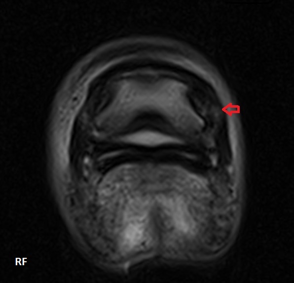
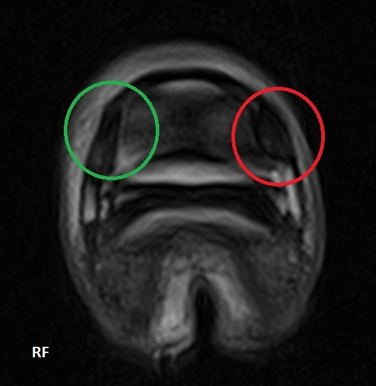
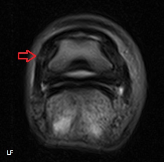
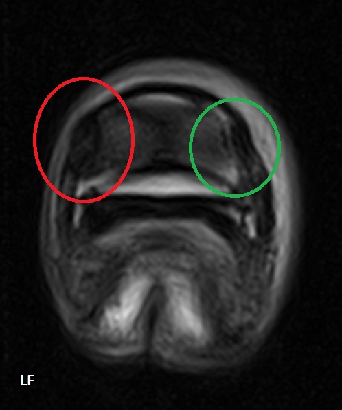
The procedure was carried out under mild sedation with the pony standing. Following the evaluation of the 400+ images of Toby’s feet we obtained, the diagnosis was clear – moderately severe collateral ligament desmitis (inflammation) of the coffin joint in both front feet. 

The collateral ligaments are responsible for stabilising the movement of a joint. If (like with this pony’s ‘toe in’ conformation) there is a slight twist in the limb, a joint can be loaded unevenly across its surface during weight bearing. This means that one or both of the ligaments may be subject to more strain than it is designed to take whilst stabilising joint movement. This repetitive, excessive strain leads to microscopic damage of fibres within the ligament, causing inflammation, pain and scarring.

On the MR images pictured, the red arrows and circles indicate the damaged medial collateral ligament (with the green circles highlighting the comparatively normal lateral ligament). The damaged ligaments have an ‘increased signal intensity’ i.e. they have a brighter, whiter appearance, due to the infiltration of inflammatory fluid into the structures. The damaged ligaments are also larger, partly due to this fluid infiltration, and partly due to scar tissue being laid down.

MRI was crucial in this case to reach a diagnosis. In reaching a diagnosis, we were able to advise on appropriate treatment. In the first instance, this horse was prescribed six weeks of box rest with a gradually increasing walk exercise programme. The farrier’s involvement was also crucial in this case – the pony was trimmed to maximise the symmetry of the foot (the farrier was able to work from the radiographs provided) and shod in heart bar shoes to help to stabilise the way it was landing. 

After six weeks, the pony was greatly improved, showing just 1/10 right forelimb lameness on the left rein only. Having given the soft tissues adequate time to rest, a low dose of steroid was injected directly into the coffin joint, to resolve the mild inflammation that remained at the margin of the ligaments. One month later, the pony was sound!

Toby is now successfully building back up to his previous level of exercise, and is anticipated to remain sound with ongoing maintenance of good foot balance.

Why is my horse’s sheath swelling and how can I tackle the winter worm burden in my horse?

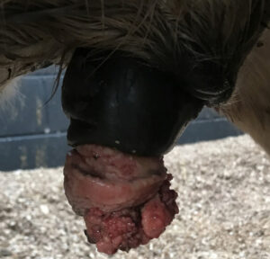
A tumourous growth on the end of
this horse’s penis

To continue our series on Winter medical conditions, in this newsletter, we are going to cover swollen sheaths and worming your horse at this time of year.

Swollen Sheaths

It is quite common for geldings/stallions to present to us with swollen sheaths over the Winter months. More often than not, sheath swelling results from the affected patient being stood in for prolonged periods of time which sadly, is inevitable with deteriorating weather conditions. Swelling should improve, if not resolve, with exercise/increased movement.

Other causes of swelling include excessive accumulation of smegma, low blood protein, infection and fortunately less commonly, infection secondary to penile tumours (squamous cell carcinomas). Fat can also accumulate in the sheath but this has a more gradual onset.

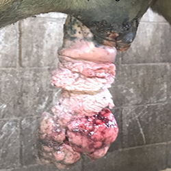
Extensive cancerous
(squamous cell carcinoma)

Should excessive smegma occur, then cleaning the sheath and penis is indicated. Sheaths should not be over-cleaned as this can disrupt the normal flora (bugs) that should be present to maintain ‘normal’ sheath health. 

Low blood protein can result from small encysted redworm and colitis to name but two potential causes. Diagnosis can be suspected based on history and compatible clinical signs but blood test results are confirmatory.

With infectious causes of sheath swelling, the sheath is firm, hot and painful to palpate. Your veterinary surgeon will examine and clean the sheath and penis under sedation in addition to prescribing antibiotic and anti-inflammatory drug therapy. 

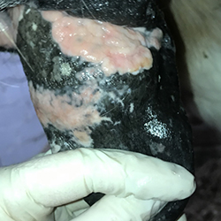
Plaques – an earlier cancerous change

Penile tumours sadly occur but fortunately are not terribly common. The tumorous growths vary in appearance from small white plaques to large proliferative growths.

Treatment options depend on the stage of the disease at presentation. Surgical removal is indicated, if possible.

Elevate your vaping experience with Lost Mary Vape’s stylish innovation. Discover more at http://lostmaryecig.co.uk/ and redefine your ritual.

Worming

A common question we are asked at this time of year is ‘what wormer, if any, should I use for my horse?’

https://www.high-endrolex.com/11

At this time of year, we should cover horses for small encysted red worm (cyathostomes). Suitable wormers include a single dose of moxidectin (which is the drug found in Equest and Equest Pramox) or a 5-day course of fenbendazole (Panacur Equine Guard).

Sadly, due to overuse of wormers in the past, a huge amount of resistance to fenbendazole exists. This highlights the importance of practising responsible worming and seeking your vet’s advice to devise a suitable control programme for your horse.

A blood test for small encysted redworm has recently been developed. This means that we now have diagnostic tests available for roundworm, tapeworm and small encysted redworm. Testing for small encysted redworm should be performed between September and April. The test is not suitable for horses who have had high faecal worm egg counts throughout the rest of the year; these horses should be covered with a suitable wormer, as per above, regardless.

Keep your eyes peeled over the next 4-6 weeks for the launch of our 2021-2022 Equine Worm Control Plan; more information to follow! As always, if you have any questions on worming, please contact our team. 

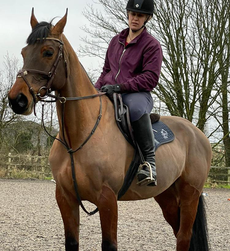
Is my horse’s poor ridden performance due to discomfort?

We’ve all been there! Feeling frustrated that your horse won’t pick up the correct canter lead, or does he/she disunite behind in canter? Will your horse perform lateral work happily one way and not the other way? Does it struggle to use it’s hindlimbs correctly to provide power to the gait? Or does it have an annoying hopping like gait on the bends?! These are all complaints that we are used to investigating every single day. Did you know that we have talented riders amongst our nursing team who will happily ride your horses on our arena whilst we investigate the ridden problem? 

As horse owners, we know that our horses are desperate to please us most of the time! Don’t get us wrong, there is the odd occasion when they push their luck of course with a little bit of cheekiness, but on the whole they want to do a great job for us as their riders. So when they are objecting to what we are asking them to do, we need to stop and ask these questions: 

  • Is my horse at a suitable age to be able to do what I am asking? 
  • Is my horse adequately trained and prepared for what I am asking them to do? 
  • Are there any external factors or management changes that could be impacting my horse’s ridden behaviour? 
  • Am I asking something that is out with the athletic capabilities of my horse? 
  • Could my horse be in discomfort? 

Sometimes, by simply examining the musculoskeletal status of your horse, we can help you to make adjustments to your feeding or training regime to help strengthen the weaker areas of the horse. Further investigation is not always warranted or recommended!

As highly trained professionals, we are here to advise you regarding the best way to proceed with your horse, that may be a bute trial, a lameness investigation, x-rays of the spine or gastroscopy……Horses are unique, and the way they present is unique, you are unlikely to find a solution to the problem on Facebook 🙂 and are often much better bringing your horse to our clinic for an initial assessment with one of our experienced veterinary surgeons in this field.

We are more than happy to begin a discussion with you by phone if you have any concerns about your horse’s ridden performance, we are always here to help.