Call Us
Goosnargh:
Lytham Road:
Kirkham:

Postpartum checks

The equine breeding season is now in full swing which means that our vets have been busy scanning mares as well attending mares and foals for routine post-partum checks as well as unfortunately, in some cases, illness.

Many of you may wonder, if the birthing process proceeds uneventfully, why the need to check mare and foal postpartum. For the remainder of this piece we will explain why this is so important in a little more detail.

Following birth, the one, two, three ‘rule’ is important to remember-

  • One: the foal should stand within one hour of birth
  • Two: the foal should show ability to nurse within two hours of birth (most mares should have expelled their placenta within this timeframe also)
  • Three: the foal should be actively nursing and consuming colostrum within three hours of birth.

If the above three timepoints are not achieved then a veterinary examination is warranted sooner rather than later. Otherwise if mare and foal are doing well, the above timepoints have been met and the mare has passed her placenta then a postpartum check should be performed within the first 24-36 hours following parturition (birthing). It is important to remove the placenta from the mare’s bed as soon as possible following expulsion and retain it for veterinary examination.

At the post-partum check, the attending vet will generally ask questions about the mare’s pregnancy and the birthing process.  A full physical examination is subsequently performed on the foal whereby the attending vet will examine the foal’s heart, chest, joints, umbilicus and check for evidence of hernias. They will also check the foal’s mouth for cleft palate and ascertain it’s jaw alignment- has it an overshot/undershot jaw. Occasionally foals have turned in eyelids which results in their eyelashes rubbing the cornea (entropion) so it is also really important to check there is no evidence of this. We will generally check the foal’s nursing also.                                                                                                        

It is highly recommended that a blood sample is taken to ascertain the foal’s IgG status. But what is this and why is it so important?

Foals are born immuno-naive which means that they are incredibly susceptible to infection should they consume inadequate quantities of colostrum (the mare’s first milk) or the mare’s colostrum is of insufficient quality. This ‘failure’ is known as ‘failure of passive transfer.’ If the mare has run milk for days on the lead up to birth, there is a distinct possibility that she will have lost the protective antibodies that would normally be concentrated in the colostrum. On the other hand, if the mare’s colostrum is of sufficient quality but the foal fails to consume sufficient quantities within the first 12 hours of life or so then the foal will consume and absorb insufficient amounts of antibodies. Both scenarios lead to failure of passive transfer. The foal’s gastrointestinal tract is capable of absorbing these antibodies most effectively during the first 12 hours of life but some absorption continues, albeit at a reduced amount, up until approximately 24 hours of age. The IgG blood test determines the foal’s antibody level. This simple blood test is performed on yard with results within a few hours at the latest. The best-case scenario is that results return indicating successful passive transfer but if test results indicate failure then the time postpartum determines the action required. If heading towards 24hrs plus postpartum, then a plasma transfusion is indicated. Your vet will discuss the most appropriate course of action with you.

It is equally as important to check the mare and her placenta as well as the foal. A full physical examination should be performed and the placenta checked. The mare’s perineal area should also be examined for birthing trauma. Finally, the mare’s bag (udder) should be checked and should be soft and well nursed. A hard, engorged bag is not normal and is often the first tell-tale sign that a foal is poorly and nursing insufficiently.

What Are Nerve Blocks & Why Are They Useful?

A nerve block is the deposition of local anaesthetic into the area immediately surrounding a nerve body or ending. This stops the nerve from transmitting signals relating to pain back to the brain, hence pain is prevented or “blocked”. Practically, the term nerve blocking is often used to describe the placement of nerve blocks in the truest sense, and also the deposition of local anaesthetic into joints and into the tissue surrounding the spinal cord (an epidural), rather than around nerves. 
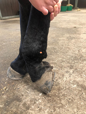
Placing needles into highly sensitive and sometimes painful areas of a horse is obviously potentially risky to the personnel involved. However experienced, calm, sympathetic handling & restraint (often coupled with some form of oral enticement!) is often sufficient to allow the placement of a block. Where this is not possible, the administration of mild sedation prior to the procedure, often allows for it to be performed safely in the vast majority of horses. 

By preventing sensation of pain, nerve blocks can be used to allow the performance of certain surgical procedures under standing sedation rather than under general anaesthetic which can decrease the risk of a procedure to the horse, and the cost to the owner. Examples of this include cheek tooth removal, sinus surgery, surgical correction of “kissing spines” (dorsal wedge ostectomy) and wound reconstruction. 

Another use is localisation of pain, most commonly orthopaedic pain or lameness – know as diagnostic analgesia. Whilst observing a lameness informs us that a horse has pain in a certain limb, it does not tell us where in the limb the problem originates. Sometimes this is obvious i.e the presence of heat, swelling and/or pain on manipulation of a certain part of the limb, but in many cases there is no visual or palpable sign of pain. It is such cases where nerve (or joint) blocking can prove invaluable. 

The procedure involves observing the horse moving in a straight line, on circles on both reins and on firm and soft surfaces. This allows grading and characterisation of the lameness. A nerve block is then placed, stopping the sensation of pain in a specific area. Once this has had chance to take effect, the horse is once again observed moving as previously. Any substantial reduction of lameness can therefore be attributed to pain coming from within the area that has been desensitised by the nerve block. If the lameness is unchanged, it can be deduced that the source of pain is not within the blocked area of the limb, and further nerve blocks can be performed to desensitise other area of the limb until the source of lameness is known. 

Once the area responsible for the lameness is known, diagnostic imaging (e.g. radiography, ultrasonography, MRI) can be performed allowing the identification of abnormalities. It is important to mention at this point, that untargeted imaging of the whole limb often results in the identification of abnormalities or variations from normal. Without having confirmed that the area being imaged is definitely the cause of lameness, it can be difficult to decide whether these abnormalities are significant 

to the lameness or just incidental findings (i.e. slight abnormalities which are present but not causing any problem). 

In conclusion, the use of nerve blocking allows identification of the region of the limb causing the lameness. This in turn allows focused imaging which results in the identification of significant abnormalities only, and a precise diagnosis. This means that the best treatment plan can be selected. It also allows an accurate and realistic prognosis to be given, i.e. how likely a full recovery is, how long it will take, and what the horse will be capable of in the future. 

The aim remaining at all times, to make your horse pain free and able to provide enjoyment to you as their dedicated owners.

Covid-19 Equine Latest

During these unprecedented times, we are closely following the guidance issued by our governing body, the Royal College of Veterinary Surgeons (RCVS). We are entirely committed to protecting the health of the public, our clients and our staff, whilst maintaining the highest possible standard of care for your horses. As of Tuesday, April 14th the RCVS have relaxed the restrictions placed on the veterinary industry. We have considered this information very carefully and whilst we will continue to provide advice on a case-by-case basis, we feel that the following guidelines contain essential information on our practice policies from April 14th:

  • We will be returning to performing annual vaccinations to horses that are due (please delay making an appointment if your annual vaccination is not due within the next 3 weeks). We will not be performing 6 monthly booster vaccinations (other than 3rd vaccines of the original course). 
  • We will continue to see any horse in an emergency situation and any horse who’s welfare is compromised. 
  • We will also be able to perform other essential veterinary work that cannot be delayed for a further 3 weeks, please ring to discuss any concerns you have with one of our vets in the first instance.
  • We will not be performing routine dentistry or other routine procedures which are non-essential.

Upon our vet’s arrival at your yard. For our mutual protection we would appreciate that the following social distancing/hygiene measures are taken:

  • We will meet one allocated person at the yard only (client), who is not self-isolating, high risk or displaying any COVID-19 symptoms. 
  • Both the vet and client will sanitise their hands and wear gloves.
  • The client will hold horses for us whilst maintaining social distancing measures, and avoiding face-to-face contact if at a distance of less than 2m (for instance as the horse is being injected).

These measures are extremely important, as the safety of our clients and our staff remain the top priority during this time. 

We are also expecting the phone lines to be busy on Tuesday morning, so please try to delay your query/appointment booking unless it is urgent.

THANK YOU ALL for being so patient and understanding during these difficult times, we are all in this together!

Our equine team and services

Oakhill Equine Vets is a 12-vet dedicated, equine-only team. We are extremely lucky at Oakhill to have a highly qualified equine veterinary team with 111 years experience between them! Guy and Rosie are both European and RCVS recognised specialists in Equine surgery and lameness. We also have two vets with RCVS advanced practitioner status, Jess and Leona. Leona has a certificate in Internal Medicine and the rest of our outstanding team are studying towards a further 7 certificate qualifications between them! 

Oakhill has a fully-equipped surgical facility including state of the art arthroscopy equipment, anaesthesia recovery box and advanced anaesthetic monitoring equipment. We host an array of specialist equipment including MRI, digital radiography, multiple endoscopes and a dental oral endoscope, in order to achieve the highest possible standards of care for your horse. 

Due to the depth of team experience and the quality of our equipment at Oakhill, we offer an extensive referral service to other practices across Lancashire, Cumbria and Cheshire. We have two vets on call 24/7 to ensure that we can attend any emergency promptly within our practice area. 

We are dedicated to providing a 5* level of service to you and your horse. Our team is also focused on providing education to clients regarding their horse’s care via phone advice, social media, informative newsletters and client talks. No matter what the problem is, we are here to offer a professional and compassionate service to our valued clients.

Laminitis

10 take home points about Laminitis

  • Laminitis is an incredibly painful and debilitating condition which, in severe cases, can result in the loss of the affected patient.
  • Laminitis is defined as inflammation of the laminae which in turn results in pedal bone instability meaning that the pedal bone may sink (founder) or rotate.
  • The primary clinical sign of laminitis is a gait abnormality which can vary vastly in severity from being foot sore or a little pottery (exacerbated on the turn), to those that are unwilling to move, to those the spend increased amounts of time lay down.
  • Other clinical signs include any or a combination of the following: increased digital pulses, heat in the feet, weight-shifting, rocking back into a ‘saw horse’ stance and sensitivity on hoof testers to name but a few.
  • Concurrent foot abscesses are a common secondary condition.
  • 90% of cases in the UK occur secondary to an underlying endocrinopathy- Equine Metabolic Syndrome (EMS) and/or Equine Cushing’s Disease.
  • Laminitis may occur at any time throughout the year although Spring turnout is a particularly ‘at risk’ time due to the sugar levels in the grass combined with many horses exiting the Winter months having gained weight and ultimately having some degree of insulin dysregulation (EMS).
  • This year, the corona virus pandemic provides further concern given the fact that many such patients are spending more time at grass or are being turned away to grass and are receiving less exercise meaning that weight gain is inevitable and the risk for EMS increased.
  • Weight management is vitally important to try prevent episodes of laminitis and may include limiting time at grass, use of a grazing muzzle, soaking hay, double netting hay and exercise provided there are no current clinical signs of laminitis.
  • If you have any concerns that your horse has clinical signs of laminitis, EMS or Cushing’s disease or if you want weight management advise, please call us for a chat. Further information on all three conditions will follow in future presentations.

Covid 19 Update

Following the Prime Minister’s announcements and advice from our professional bodies, we are moving to a service where we no longer provide any ROUTINE veterinary care. This is to ensure the health and safety of our staff and our clients, and to strictly adhere to social distancing policies as set out by the government.

We will be providing care to ANY ill or injured animal in order to preserve animal welfare. Please contact the surgery as normal with any concerns you have regarding your horses’ welfare. We may only need to offer phone advice, and any horses that require treatment in order to preserve welfare will be examined. These decisions will be made on a case by case basis and the handler of the horse will need to confirm they are free of COVID 19 symptoms prior to our attendance.

We will continue to offer our normal 24 hour emergency cover. Your horse’s welfare is paramount to us and we thank you for your understanding at this difficult time. If you have any questions or would like to speak to a vet, please contact the office on 01772 861300

Equine Standing MRI – what to expect

What to expect when your horse has a standing MRI.

Using the right tools early in the lameness process to get a definitive diagnosis will allow you and your vet to devise the right treatment plan, hopefully getting your horse back to full fitness as quickly as possible.

The clear images from MRI allow vets to make an accurate and precise diagnosis in 90% of cases.

If you’ve considered requesting an MRI for your horse but wondered what actually happens, an MRI scan will usually include the following steps:

  1. Initial examination

    On arrival for the scan the horse’s overall health is evaluated for sedation and our clinic vets will briefly examine the horse’s lameness.


  2. Horse shoes

    Metal horse shoes would degrade the quality of the images if left on as the MRI scanner contains a large magnet. Normally just two shoes, on the leg to be scanned and the adjacent leg, are removed.


  3. Sedation

    The standing MRI eliminates the need for anaesthesia, so removes the mortality risk and often allows for day patient scheduling. Top up doses may be applied during the scan, either on a drip or via a catheter in the horse’s jugular vein.


  4. Equine Standing MRIPositioning

    The horse is walked into the MRI scanner, with the lame leg placed between the poles of the magnet. A radiofrequency coil is fitted around the injury site and the operator makes careful adjustments to ensure the horse and magnet are both in the right place.


  5. The Scan

    equine MRIThe scan takes around 2 – 4 hours, producing around 300-500 images at multiple angles of the limb or hoof, highlighting different types of tissue and pathology.


  6. Recovery

    After the scan the horse is given time to recover from the sedation, and in most cases can return home the same day.


  7. Interpretation

    One of our specialists responsible for scanning will carefully review the images to arrive at an opinion about likely pathology or injury. The findings are then communicated to you or your vet.


  8. Treatment

    The findings from the scan will enable an accurate diagnosis to be made. With precise information available the vet can prescribe the best possible treatment for the horse.

Should your horse be suffering with lameness or poor performance issues, please discuss with your usual Oakhill Equine Vet or call the practice on 01772 861300.

If you wish to be referred to us from another veterinary practice, please contact your own veterinary surgeon in the first instance.

Why MRI?

Our equine clinic has the most technologically advanced standing MRI machine available, used for both clients and those referred from other veterinary practices.

Often during a lameness work-up, your vet will use ‘nerve blocks’ to locate where the pain is coming from. This may then be followed with X-ray or ultrasound examinations, but because these imaging tools only show bone or some soft tissue your vet may not be able to see abnormalities, particularly those within the hoof capsule.

Standing equine MRI offers unique insight into equine lameness, identifying the specific cause in over 90% of cases.

With no general anaesthesia required the inherent risks associated with anaesthesia and recovery can be avoided and an early, safe and accurate lameness diagnosis will save you time, money and worry.


We’re often asked….

equine MRIWhat is different about MRI?

MRI images show information about both bone and soft tissue, whereas x-rays only show bone and ultrasound only shows soft tissues. The many, clear images from MRI allow vets to make an accurate and precise diagnosis in 90% of cases.

Foot Lameness Cycle


Oakhill equine MRI referral serviceIs it the same as a human MRI scanner?

The underlying principle is exactly the same but the Hallmarq Standing Equine MRI system has specifically designed for imaging the standing horse, not a human! The scanner operates close to floor level and the horse can immediately step out of an opening in the magnet if it needs to.


Is it safe for my horse?

MRI is widely used in both human and veterinary medicine as it is valued for it’s high image quality of both bone and soft tissue with no ionising radiation. The technology used is unobtrusive and poses no known risk to the horse.

Unlike high-field scanners where the horse has to be anaesthetised, our Hallmarq low-field system uses a smaller magnet that fits around the leg, allowing us to image the horse while standing and under mild sedation.

Hallmarq equine MRI systems have been used for over 60,000 standing sedated horse examinations, during this time there have been no fatalities.


The benefits of having an MRI scan at Oakhill Equine Clinic include:

  • Expert interpretation of images by our ECVS & RCVS recognised surgery specialists, Rosie Owen & Guy Hinnigan and imaging specialist, Meredith Smith.

  • Precise diagnosis

  • Rapid results

  • Specific prognosis

  • Optimum treatment

Using the right tools early in the lameness process to get a definitive diagnosis will allow you and your vet to devise the right treatment plan, therefore getting your horse back to full fitness as quickly as possible.

It is also very useful in establishing an accurate prognosis, to save you time and money worrying about what is likely to happen in the future.

Click here to find out more about what to expect when your horse has a standing MRI.

Should your horse be suffering with lameness or poor performance issues, please discuss with your usual Oakhill Equine Vet or call the practice on 01772 861300.

If you wish to be referred to us from another veterinary practice, please contact your vet in the first instance.

Mission Christmas Gift Appeal

Once again Oakhill Veterinary Centre will be taking part in Rock FM’s Mission Christmas Gift Appeal 2019, in aid of Cash for Kids, who make sure Christmas gifts get to disadvantaged children in the area.

All 4 of our branches will be gift collection points.

If you’d like to donate a gift, it should be new and unwrapped (no food or chocolate please) and be with us by Monday 16th December at the latest so we can get them to Mission Christmas HQ in time.

Further information is available on the link below, including gift ideas (from birth to 18 years):

https://planetradio.co.uk/rock-fm/charity/events/mission-christmas-lancashire/

Thank you!

Keeping your horse safe on Bonfire Night

Bonfire Night can be stressful for both horses and horse owners.

The calmest of horses can become frightened by the unfamiliar loud noises and flashes of bright light.

There are some things you can do to prepare….

  • Find out when and where the firework displays will be in your area. You could look in local press, social media and shop notice boards.
  • Inform local firework display organisers and neighbours that there are horses nearby, so they can ensure fireworks are set off well away from them.
  • Decide whether to stable your horse or leave it in the field. Horses like routine, so try and keep as close to your usual routine as possible. If it is usually stabled, keep it stabled. If it is normally out in the field, keep it there as long as it is safe, secure and not close to the firework display area.
  • Check for anything that could cause injury to your horse:
    – If stabled, look for things such as protruding nails.
    – If your horse is to stay in the field, check that fencing is not broken and that there are no foreign objects lying around.
  • Ensure that you, or someone experienced, stays with your horse if you know that fireworks are being set off.
    If you have to leave your horse in the care of another person during a firework display, then be sure to leave clear instructions with contact details for both you and your vet.
  • Discuss with your vet about sedation or perhaps consider moving your horse for the night.
  • Playing music on a radio positioned outside the stable can often mask sudden noise, distract attention and be soothing.
  • Stay calm, as horses will sense that you are anxious and that could increase their fears.
  • A startled horse can be dangerous, so try not to get in the way if a horse becomes frightened as you could easily be injured yourself.
  • Remember that Chinese lanterns (also known as sky lanterns) can cause harm to livestock and wildlife.

If you wish to discuss concerns about your horse with us, please call 01772 861300