Call Us
Goosnargh:
Lytham Road:
Kirkham:

Keeping your horse safe on Bonfire Night

Bonfire Night can be stressful for both horses and horse owners.

The calmest of horses can become frightened by the unfamiliar loud noises and flashes of bright light.

There are some things you can do to prepare….

  • Find out when and where the firework displays will be in your area. You could look in local press, social media and shop notice boards.
  • Inform local firework display organisers and neighbours that there are horses nearby, so they can ensure fireworks are set off well away from them.
  • Decide whether to stable your horse or leave it in the field. Horses like routine, so try and keep as close to your usual routine as possible. If it is usually stabled, keep it stabled. If it is normally out in the field, keep it there as long as it is safe, secure and not close to the firework display area.
  • Check for anything that could cause injury to your horse:
    – If stabled, look for things such as protruding nails.
    – If your horse is to stay in the field, check that fencing is not broken and that there are no foreign objects lying around.
  • Ensure that you, or someone experienced, stays with your horse if you know that fireworks are being set off.
    If you have to leave your horse in the care of another person during a firework display, then be sure to leave clear instructions with contact details for both you and your vet.
  • Discuss with your vet about sedation or perhaps consider moving your horse for the night.
  • Playing music on a radio positioned outside the stable can often mask sudden noise, distract attention and be soothing.
  • Stay calm, as horses will sense that you are anxious and that could increase their fears.
  • A startled horse can be dangerous, so try not to get in the way if a horse becomes frightened as you could easily be injured yourself.
  • Remember that Chinese lanterns (also known as sky lanterns) can cause harm to livestock and wildlife.

If you wish to discuss concerns about your horse with us, please call 01772 861300

Osteoarthritis in the adult horse

Osteoarthritis (OA) is one of the most common causes of lameness in the adult horse. It is primarily a disease of the articular cartilage, a thin layer of protective material covering the surface of bones within joints, with progressive deterioration which can result in pain and inflammation.

Whilst OA can be a significant source of pain, especially when the horse is being worked, this can often be controlled and horses are often able to continue being ridden and even competing if well controlled.

There are many factors which may influence the development of OA in a horse- most commonly it is thought of as a wear and tear condition of older horses following years of hard work, but it can also develop suddenly in younger horses as a result of developmental joint disease (DJD), trauma or injury. Other compounding factors include conformation, weight and genetics.

The diagnosis of OA is often made following a multimodal approach by your vet. This normally starts with a clinical examination, in which the vet will palpate and manipulate the limbs with the horse stood still, and then watch it move in a straight line and on a hard and soft lunge circle. Flexion tests are then often performed to increase the pressure on the joints and surrounding soft tissue structures. The next stage is usually nerve-blocking with local anaesthetic; this allows the area of pain to be identified by removing the sensation to different regions of the limb. A positive nerve block will often result in abolition of the lameness when the dynamic tests are repeated and an improvement in the horse’s way of going. The local anaesthetic can either be placed under the skin next to a nerve (perineural) or into a joint (intra-articular).

Following this, imaging of the joints is normally required with radiography (x-ray) the main technique used. This allows assessment of the bones of the joint including their density, shape and the space around them.

Radiographic changes frequently observed with OA may include new bone formation or bone loss, reduction of joint space and irregular margins. Other imaging modalities include ultrasonography, nuclear scintigraphy and even magnetic resonance imaging (MRI), however these are more often used in unusual or complex cases of lameness originating from more than one source or in a horse not amenable or with inconclusive nerve blocking.
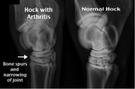
Osteoarthritis radiograph

Once OA of a joint has been diagnosed the treatment is usually conservative. Each case will be considered as an individual, with the aforementioned compounding factors and the horse’s workload and work history playing an important part in the management plan. Initial treatment may involve a combination of physical and drug-based approaches.

Early diagnosis can be very beneficial to long-term management by implementing changes which may slow the progression of disease, alleviate pain and maintain the horses’ way of going. Concurrent work with your physiotherapist and farrier may also help by providing additional musculoskeletal support. Gentle ongoing low-grade movement is usually beneficial with inactivity causing further stiffening so daily exercise or turn out is important.

Pharmacologically, the mainstay treatment is oral non-steroidal anti-inflammatory (NSAID) drugs such as phenylbutazone (bute) or suxibuzone (danilon). Whilst they will not modify the course of the disease, they will provide pain relief and reduce inflammation which in turn will allow the horse to keep moving. Long term these may have side effects including kidney and gastro-intestinal issues and such use should therefore be monitored and is often cautioned against. Alternative pharmacological intervention includes direct corticosteroid medication into the joint. This provides a targeted anti-inflammatory effect and will also have the potential to modify the disease process. However used incorrectly or over frequently, joint medication may have deleterious effects on the joint cartilage as well, and also carry the potential increased risk of laminitis and joint infection in the acute short-term.

Other treatment options include the use of bisphosphates, hyaluronic acid, and joint supplements (neutraceuticals). There is limited scientific evidence to support the use of neutraceuticals in horses, although recent studies have highlighted boswellia and green-lipped muscles as potentially important ingredients. A last-resort treatment suitable to some joints is surgical or chemical fusion (arthrodesis). This reduces the potential for movement in a low-motion joint and can be very effective at alleviating pain, although is an irreversible change and may not be suitable for many cases.

A further area of increasing interest is the use of biological therapies; i.e. re-introducing cells or proteins extracted from a horse, back into the damaged area or joint. Currently these include PRP and IRAP, and are designed to increase the health of the joint fluid and stimulate cellular repair. A new approach that is increasingly used is a polyacrylamide hydrogel with can be injected into some joints to increase cushioning and thus alleviate pain.

It is important to weigh up the safety, proven efficacy and cost of any of these treatments, and what may be of most benefit to your horse. There are no medical treatments that can reverse the cartilage damage associated with OA. The best approach is to find a balance of management for the individual which will suit you both as a team.

If you’d like to discuss with a member of the Oakhill Equine Vet Team, please contact us.

Equine Castration Clinics

Equine Castration Clinics

Monday 29th April 2019 & Thursday 23rd May 2019

at the Oakhill Equine Clinic, Langley Lane, Goosnargh, Lancashire, PR3 2JQ

Only £130 + VAT

Price includes: Procedure under general anaesthesia, sedative, pain relief and antibiotics.

If horse is not vaccinated, an additional cost will be added for tetanus anti-serum.

Call 01772 861300 to book.

Terms and Conditions

Horse must be under 3 years old.
Offer only applicable on dates shown and to be carried out at Oakhill Veterinary Centre, Langley Lane, Goosnargh, PR3 2JQ.

Price is per horse. Payment must be made on collection of your horse. Passport needs to be presented and the horse to be certified as ‘not for human consumption’ to enable the relevant drugs to be administered during procedure.

The procedure cost does not include the cost of treatment as a result of any post-operative complications which may arise.

If one or both testicles are not descended a different type of surgery will be required – this will need to be arranged separately and additional cost will be charged.

Laminitis

As the days get longer and the sun is out, horses will be enjoying the perks of being turned out more. This could be a potential danger for horses at risk of laminitis or overweight.

Recent research has shown that laminitis is as common in horses in the UK as colic. In contradiction to what was previously thought, there is no safe-season for laminitis, and it is presented and diagnosed all year around¹.

It is important that owners remain proactive about the prevention of laminitis in every season of the year. The earlier an episode is recognised significantly increases the best chance of recovery. This includes looking out for the perhaps more subtle signs, including changes in behaviour, reluctance to pick up feet, a shortened/stiffened gait and being careful on hard stony grounds.

Laminitis

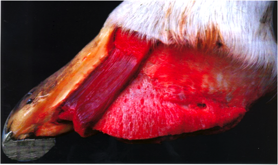
Picture 1. Hoof wall with laminae dissected.

The horse’s foot is built from a sensitive (soft tissue inside) and a non-sensitive part (hoof wall). The sensitive parts are called the laminae, which connect the hoof wall to the pedal bone inside (Picture 1).

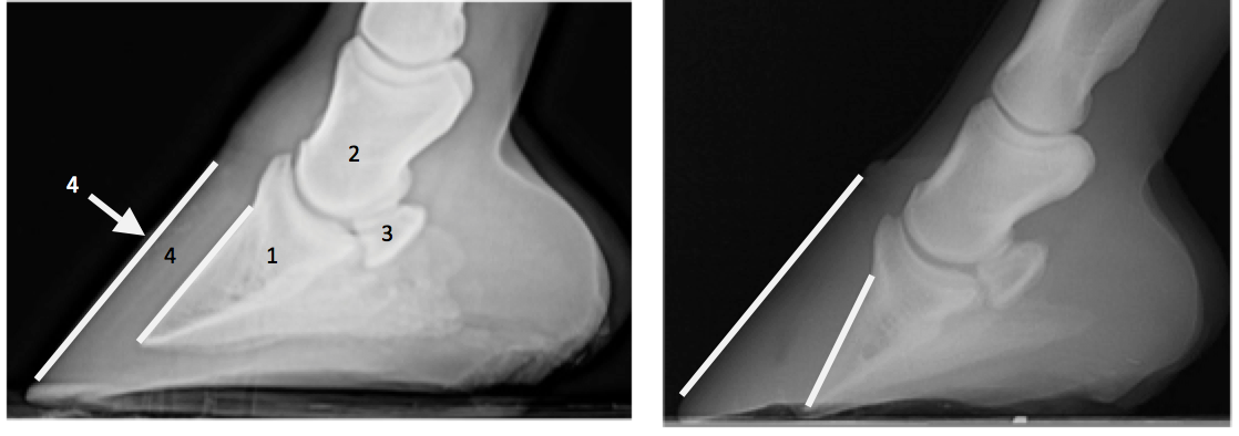
In a horse with laminitis these laminae get inflamed, causing them to loosen the connection. Because of this the pedal bone will rotate towards the sole of the foot. The inflammation and rotation of the pedal bone are very painful for the horse. The rotation of the pedal bone can only be confirmed by taking a xray (Picture 2).

Laminitis Xray

Picture 2.

  1. Pedal bone  2. Short pastern bone. 3. Navicular bone.  4. Laminae

Left xray shows normal anatomy with the pedal bone parallel to the hoof wall, right xray shows a rotated pedal bone, where the tip of the pedal bone has penetrated the sole.

Restricted grazing in horses at risk of being overweight and laminitis is indicated, ideally horses at risk should not be turned out on grass at all.

Strip grazing, limiting the hours of turn out on grass and turning out with a muzzle are advised, but some horses will adapt to this.

If you think your horse is overweight, please don’t hesitate to book yourself onto one of our weight clinics!

¹Pollard et al. (2018) Incidence and clinical signs of owner-reported equine laminitis in a cohort of horses and ponies in Great Britain

Foaling season

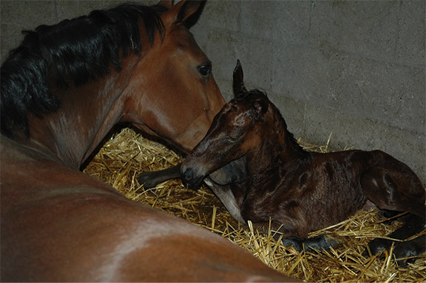
Exciting times if your mare is about to foal this season!

A couple of days before parturition the udder of the mare will develop wax tips on the end of the teats.

A normal birth usually takes about 30 minutes without any help. Directly after the foal is born the umbilicus is still attached. It is important to leave the mare and foal attached for as long as possible. The umbilicus will break at the right time and place, there is no need to cut it. The first couple of hours are important for the foal to drink enough colostrum. Colostrum is full of antibodies necessary for the immunity of the foal.
Within 8 hours the foals should drink approximately 2-3 litres of colostrum to get a sufficient enough antibodies.

The foal is only able to take up any antibodies in the first 24hours. If there are any concerns about the uptake of colostrum, a blood sample can be taken to test for IgG levels.

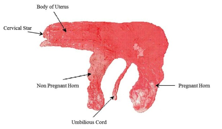
The placenta should be detached from the mare within 4 hours after parturition, it is important to check if the placenta is complete (needs to look like a pair of trousers). If the placenta has not come out within 4 hours, or is not complete it is important to contact the vet.
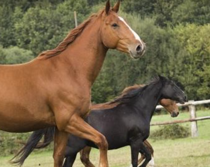
Did you know that a healthy foal should:

[checklist icon=”fa-arrow-circle-right” iconcolor=”#39aa87″ circle=”yes”]

  • Lift its head < 5 min

  • Stand < 2 hours

  • Drink < 2 hours

  • Urinate < 6-10 hours

  • Pass yellow feces < 24 hours (after dark meconium)

[/checklist]

It is advisable to have your mare and foal checked 24 hours after foaling, even if everything went smoothly. Foals are very good at fooling us! If the foal is a bit dull and quiet, not drinking enough or any other concerns you should ring your vet immediately. In these cases the earlier the foal is seen the better.

Diastemata

The 24 cheek teeth are designed to grind forage for up to 16 hours a day. These teeth erupt and are ground down continuously and those in each quadrant of the mouth act as a single grinding surface without spaces.

A diastema (plural, diastemata) is defined as a gap between teeth. Diastemata in the normally tightly fitting cheek teeth results in food entrapment. The stagnant food leads to painful periodontal disease (inflammation of the gums). If left untreated this can lead to the loosening of teeth, tooth root infections, sinusitis or food tracking up into the nose.

40-50% of horses suffer from diastemata. They are most common in older horses. It’s our most commonly treated dental disease.

Diastemata can result for many reasons:DIASTEMA

[checklist icon=”fa-arrow-circle-right” iconcolor=”#39aa87″ circle=”yes”]

  • With older age, teeth have erupted to the level of the slightly narrower root portion of the tooth.
  • Displaced (out of line) or rotated teeth- both are developmental defects, more common in breeds with small or dished faces e.g. miniatures.
  • Teeth drifting apart, towards the space remaining following a tooth extraction.
  • The teeth have erupted with diastemata between them, perhaps because the horse had grown bigger than the relative size of its teeth or erupted with out angulation.
  • Caries (decay) of the teeth which rots tooth away creating diastemata between them.

[/checklist]

Periodontal disease is very painful and horses often present with quidding (dropping of balls of hay). Some horses ‘pull faces’. Subtle signs e.g. eating less or weight loss are often attributed to old age. Symptoms are commonly seen during winter as long forage is more likely to become trapped between teeth than grass.

Diastemata are diagnosed at a routine oral exam, recommended every 6-12 months. To perform a thorough examination, a headtorch and mirror is essential; diastemata can be easily missed. Sedation is useful for fidgeting horses.

Treatment is carried out using sedation, pain relief and local anaesthetic in our clinic and involves removal of trapped food with the guidance of the oral endoscope camera that can view into the deepest pockets. Widening of diastemata is sometimes required to release entrapped food. The cleaned deeper pockets are sometimes packed with a protective putty whilst the gum heals beneath. Tooth overgrowths are floated (reduced) using powertools in order to balance the pressures on the teeth, reducing tooth drift. Severe cases may require tooth extractions.

Following a course of thoroughly performed treatment, the prognosis for resolution is very good especially with early diagnosis and action.

Equine Influenza – what’s the current situation?

There have been separate cases of Equine Influenza confirmed in the UK. Horses in Essex, Cheshire and Derbyshire have been affected this month with further reports of outbreaks in Belgium, France and Germany. With all three outbreaks it has been unvaccinated horses that have tested positive.
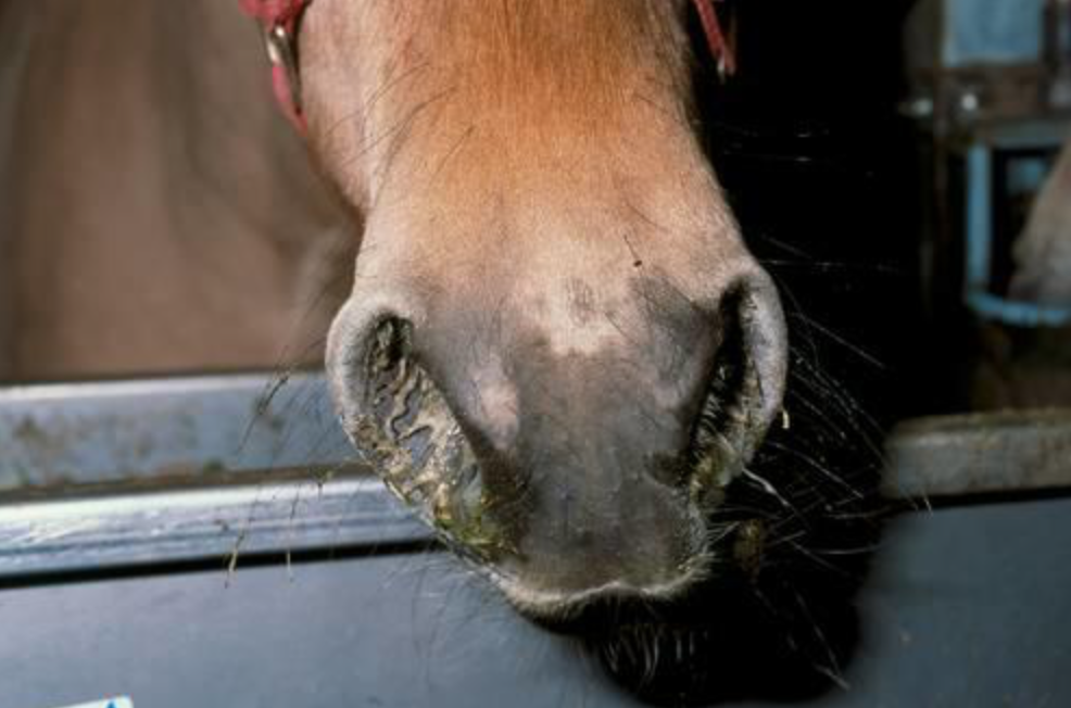
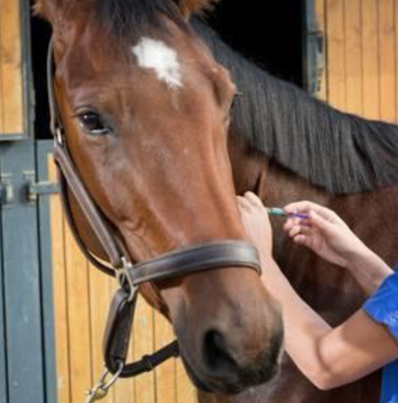
Currently the number of outbreaks is small but they have occurred within a short duration; to put things into perspective a similar number of cases were reported during the whole of 2018.

It is currently unknown which strain of the virus has caused these outbreaks, but research is being conducted to find out. While there are several signs of flu in horses, those affected may only show one or two of them.

What are the signs of infection?

Unvaccinated horses

  • In unvaccinated horses the virus targets the upper respiratory tract, this leads to a very dry, harsh or hacking cough.
  • They usually have a raised temperature which can last around 7-10 days. The fever often makes them dull, off their food and lethargic
  • A clear or white nasal discharge is often seen with enlargement of the submandibular lymph nodes (under their chin/throat area).

Vaccinated horses

  • Horses that have been regularly vaccinated often show no clinical signs, but they may still shed enough virus to infect other horses. This is how the outbreak in Australia in 2007 started.
  • Horses that have been vaccinated but only have partial protection, e.g. because they haven’t been vaccinated frequently enough or because the vaccine type used was not updated, may show varied signs of mild non specific respiratory disease. This can include mild lethargy, nasal discharge and possibly a cough.

What to do if you think there is a possible infected horse?

Owners should seek veterinary advice if they suspect there is flu on the yard and carry out the following precautions:

  • Isolate the possibly infected horses. Infected horses will spread the virus in respiratory droplets, through coughing, and can spread the virus for up to 10 days. They must be isolated until the vet advises they are no longer infected.
  • All horse movements on and off the yard should be stopped.
  • Monitor all horses on the yard for clinical signs and record their rectal temperature daily, it should be less than 38.0˚C. A rise in temperature can be an early sign of an infection.
  • Booster vaccinating all in-contact horses, even if they are not yet due their annual booster, has been shown to provide horses with even more protection against flu.

How to prevent it

Horse Flu is endemic in the UK, which means that most horses will come into regular contact with the virus during their lifetime. The control of horse flu in the UK is based on limiting the signs of infection in horses that have been exposed to the virus, rather than trying to prevent exposure itself. This is primarily achieved through regular vaccination which is given either once or twice a year.
If you have any questions about your horse’s vaccinations, please call our Equine Team on 01772 861300

Free* ACTH tests are back for 2019

Testing for PPID in your horse or pony

A few years ago PPID or Equine Cushing’s Disease was considered a rare hormonal disease in horses. Now it is thought to affect over 20% of horses over the age of 15 and is a condition recognised almost daily in equine veterinary practice.

Past ‘Talk about Laminitis’ test results show that you should look out for any of these clinical signs in your horse:

  • Laminitis
  • Abnormal or delayed moulting
  • Muscle wastage
  • Periorbital fat (fat round the eyes)
  • Increased drinking and urinating

In 2017, thousands of laminitic patients benefitted from PPID being ruled in or out of their condition during the Talk About Laminitis campaign, as a result, the Talk About Laminitis campaign will continue offering FREE* ACTH lab testing during 2019

You can claim your complimentary testing voucher code by visiting www.careaboutcushings.co.uk

If your horse is already on Prascend, you may be eligible for a free monitoring test (lab fees only). You can generate your voucher code for this by visiting the link above.


Terms & Conditions:

  • This free ACTH test voucher code is for the laboratory fees for a blood ACTH test only.
  • Each voucher is only redeemable against an ACTH blood test where the patient has not been previously diagnosed with PPID
  • Full T&Cs can be found below at www.careaboutcushings.co.uk
  • Oakhill blood sampling fee, visit fee and postage still apply.

What lurks beneath – equine lice

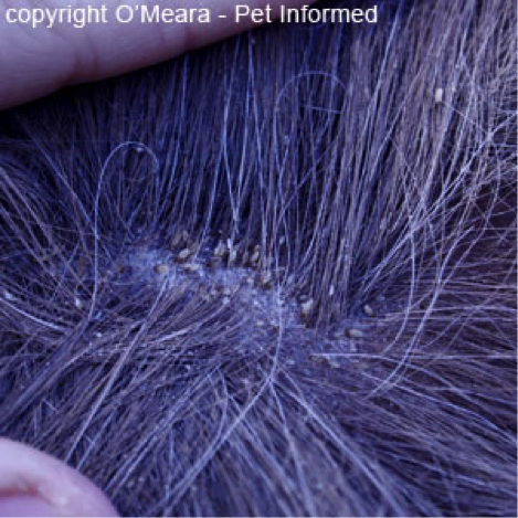
As our horses grow a thicker and longer coat over winter to keep themselves insulated it becomes an ideal breeding ground for lice, especially when we then cover this in a warm rug.

The life cycle of the louse is complete within 4 weeks and eggs hatch 10 days after being laid. Eggs or nits are small (1mm), yellowy-white in colour and glued tightly to the base of hairs.

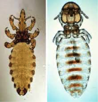
Damalinia

Haematopinus Lice

Haematopinus

All horses can become infected with lice and those kept in large groups indoors, the elderly and young and those with immunosupressive conditions such as malnourishment and PPID (Cushings) are at an increased risk.

There are two types of lice affecting horses, a biting louse called Damalinia equi and a sucking louse called Haematopinus Asini.

Biting lice feed on skin scales and are found where the coat is finer (neck & body) whereas sucking lice feed on blood and are found where the hairs are longer in the mane and tail.

The feeding nature of the lice causes skin irritation with horses becoming extremely itchy. This can lead to patchy hair loss and a scurfy coat. In severe infestations the horse or pony can become anaemic.

Diagnosis is based on evidence and appearance of lice or their eggs.

Removal of the winter coat will help ensure treatment reaches the skin and has the best effect. Lice powders are available over the counter however anecdotally they don’t seem to work. Treatment needs to involve a permethrin or cypermethrin product applied every two weeks for a few treatments to ensure all life stages of the lice are killed. In addition if there is an underlying cause this needs to be identified and treated to prevent reoccurrence.

The lice affecting horses can’t affect humans however sharing rugs/ numnahs/ grooming kits can spread it horse to horse.

If you have any questions regarding lice on your horse, please contact us.

Winter Series: Mud Fever

Mud fever, also known as greasy heel syndrome but correctly termed pastern dermatitis, is an infection of the skin usually of the pastern.

The bacteria causing the infection can be from the environment or living on the skin itself and will gain entry due to an injury to the skin surface. It more commonly affects white socked legs, as the skin’s immune function of these areas is reduced. In minor cases it may present as just a few scabs, however, it can spread above the fetlock, occur concurrently with cellulitis, or leg filling, and be openly bleeding.

Although it can have a typical appearance sometimes a vet visit is needed to rule out other conditions and to identify predisposing factors.

The skins integrity is weakened by continuous wetting of the skin and rubbing from mud meaning it is a problem commonly seen in Winter. However, it may not just be wet and muddy conditions that are causing the problem……..

[checklist icon=”fa-arrow-circle-right” iconcolor=”#39aa87″ circle=”yes”]

  • Lymphoedema: this is typical of cob type or draught breeds. Lymph fluid circulates around the body carrying nutrients in lymph vessels. The lymph vessels in these types seem to be less effective in circulating blood in the lower limbs which leads to accumulation of fluid and skin thickening.
  • Leucocytoclastic vasculitis: this is an immune mediated condition whereby blood vessels become damaged. Personal experience shows it occurs more commonly on the outside of white pasterns.
  • Photosensitisation: this is caused by either liver disease or allergic reaction meaning the horse doesn’t deal with chemicals that react with light meaning that when the horse is exposed to UV radiation (sunlight) it becomes sunburnt.
  • Feather mites/ Chorioptic mange: Chorioptes mites causes itchiness of the lower limbs. This can lead to self trauma and wounds allowing skin infection.

[/checklist]

To rule out these conditions it may be necessary to take blood samples or biopsies to determine if one of these are involved.

In order to treat Mud Fever you may need to treat the underlying cause. Sometimes a thorough antibacterial cleaning and removal of scabs is needed, under sedation, so that topical antibacterial, steroidal and protectant creams can be applied directly onto the damaged skin. In some cases when associated with cellulitis or more widespread infection of the leg systemic antibiotics may be needed.

The deliberation is always; do you wash the mud off your horses legs or do you brush it off when dry ?? In our opinion neither is superior. If you are to wash your horses legs they need to be thoroughly dried and if there is already some mud fever present avoid wetting the leg further.

If you are concerned about Mud Fever in your horse, please call us on 01772 861300