Call Us
Goosnargh:
Lytham Road:
Kirkham:

Foaling season

Exciting times if your mare is about to foal this season!

A couple of days before parturition the udder of the mare will develop wax tips on the end of the teats.
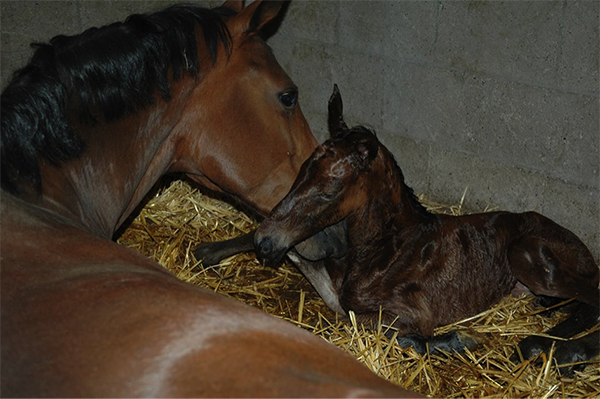
A normal birth usually takes about 30 minutes without any help. Directly after the foal is born the umbilicus is still attached. It is important to leave the mare and foal attached for as long as possible. The umbilicus will break at the right time and place, there is no need to cut it. The first couple of hours are important for the foal to drink enough colostrum. Colostrum is full of antibodies necessary for the immunity of the foal.

Within 8 hours the foals should drink approximately 2-3 litres of colostrum to get a sufficient enough antibodies.

The foal is only able to take up any antibodies in the first 24hours. If there are any concerns about the uptake of colostrum, a blood sample can be taken to test for IgG levels.
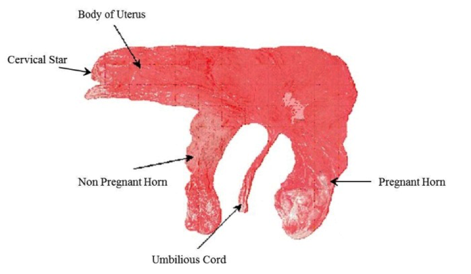
The placenta should be detached from the mare within 4 hours after parturition, it is important to check if the placenta is complete (needs to look like a pair of trousers). If the placenta has not come out within 4 hours, or is not complete it is important to contact the vet.

Did you know that a healthy foal should:

[checklist icon=”fa-arrow-circle-right” iconcolor=”#39aa87″ circle=”yes”]

  • Lift its head < 5 min

  • Stand < 2 hours

  • Drink < 2 hours

  • Urinate < 6-10 hours

  • Pass yellow feces < 24 hours (after dark meconium)

[/checklist]

It is advisable to have your mare and foal checked 24 hours after foaling, even if everything went smoothly. Foals are very good at fooling us! If the foal is a bit dull and quiet, not drinking enough or any other concerns you should ring your vet immediately. In these cases the earlier the foal is seen the better.

Diastemata

The 24 cheek teeth are designed to grind forage for up to 16 hours a day. These teeth erupt and are ground down continuously and those in each quadrant of the mouth act as a single grinding surface without spaces.

A diastema (plural, diastemata) is defined as a gap between teeth. Diastemata in the normally tightly fitting cheek teeth results in food entrapment. The stagnant food leads to painful periodontal disease (inflammation of the gums). If left untreated this can lead to the loosening of teeth, tooth root infections, sinusitis or food tracking up into the nose.

40-50% of horses suffer from diastemata. They are most common in older horses. It’s our most commonly treated dental disease.

Diastemata can result for many reasons:DIASTEMA

[checklist icon=”fa-arrow-circle-right” iconcolor=”#39aa87″ circle=”yes”]

  • With older age, teeth have erupted to the level of the slightly narrower root portion of the tooth.
  • Displaced (out of line) or rotated teeth- both are developmental defects, more common in breeds with small or dished faces e.g. miniatures.
  • Teeth drifting apart, towards the space remaining following a tooth extraction.
  • The teeth have erupted with diastemata between them, perhaps because the horse had grown bigger than the relative size of its teeth or erupted with out angulation.
  • Caries (decay) of the teeth which rots tooth away creating diastemata between them.

[/checklist]

Periodontal disease is very painful and horses often present with quidding (dropping of balls of hay). Some horses ‘pull faces’. Subtle signs e.g. eating less or weight loss are often attributed to old age. Symptoms are commonly seen during winter as long forage is more likely to become trapped between teeth than grass.

Diastemata are diagnosed at a routine oral exam, recommended every 6-12 months. To perform a thorough examination, a headtorch and mirror is essential; diastemata can be easily missed. Sedation is useful for fidgeting horses.

Treatment is carried out using sedation, pain relief and local anaesthetic in our clinic and involves removal of trapped food with the guidance of the oral endoscope camera that can view into the deepest pockets. Widening of diastemata is sometimes required to release entrapped food. The cleaned deeper pockets are sometimes packed with a protective putty whilst the gum heals beneath. Tooth overgrowths are floated (reduced) using powertools in order to balance the pressures on the teeth, reducing tooth drift. Severe cases may require tooth extractions.

Following a course of thoroughly performed treatment, the prognosis for resolution is very good especially with early diagnosis and action.

Neonatal Lamb Care

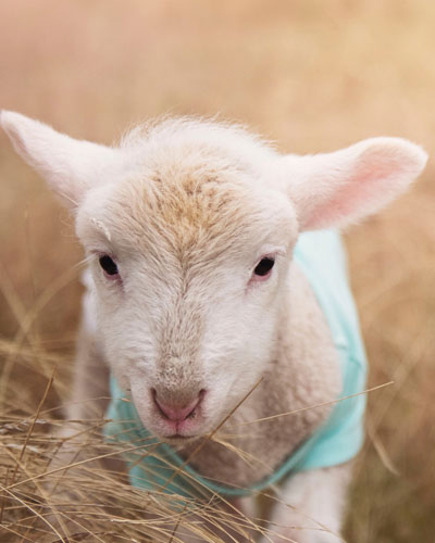
Neonatal Lamb CareLambing time is the most crucial part of the year for making your sheep business profitable. Lamb deaths from birth to 3 days old should be less than 7% however many farms range between 10- 25%.
A major factor influencing mortality in neonate (young) lambs is ewe body condition; as this directly relates to lamb body weight, colostrum quality and quantity. It is therefore important to ensure ewes are at BCS 2.5 to 3.0, (so vertebral spinous processes only felt with pressure and transverse processes only felt with hard pressure), and they are on a rising plane of nutrition in the run up to lambing. Energy and protein blood profiles can be taken 2 to 3 weeks before lambing to ensure ewes are receiving the correct nutrition to help reduce cases of twin lamb disease.

Another major cause of neonatal mortality is watery mouth. These lambs typically are lethargic, salivate and have a distended abdomen. It is caused by Ecoli and picked up by ingestion. Treatment involves electrolytes and antibiotics but is often hopeless. Therefore the emphasis should be on prevention which involves keeping pens clean, (especially towards the end of the season), dipping navels with strong iodine and ensuring all lambs get enough good quality colostrum quickly. See below:


Always ensure colostrum is…

QUALITY

Ensure ewes in good BCS, if you’re uncertain as to the quality of colostrum; colostrum quality can be measured using a refractometer: aim for specific gravity >1.05. Only use sheep colostrum replacement (colostrum from a milking sheep farm will usually be better quality than powdered colostrum); cow colostrum can cause a fatal anaemia. Also remember to give your Clostridial and Pasteurella vaccine four to six weeks before lambing to ensure the lambs receive antibodies against these diseases. (Any ewes that haven’t had this vaccine before will need a course of two injections four to six weeks apart).


QUANTITY

200ml/kg within 18 hours of birth, with a maximum of 50ml/kg intake on each occasion.

Remember lambs in adverse weather conditions will require more colostrum or milk as more energy from their feed will be used to keep them warm.


QUICKLY

Within 2 hours of birth.


SQUEAKY CLEAN

Ensure ewe udders are clean and dry and the equipment to mix and administer colostrum is cleaned and disinfected between lambs.


QUANTIFY

We can also look if your lambs have received enough colostrum by blood testing any less than a week old.


Finally, there is continual pressure on antibiotic use. So to ensure we don’t end up with a world full of super bugs we need to be careful how we use antibiotics. Preventative use of antibiotics, such as blanket use of Spectam or Betamox LA for watery mouth needs to be reduced as this will actively select for resistance and there is pressure from Red Tractor Assurance to reduce this practice.

We recommend only using antibiotics….

    • Towards the end of the lambing season, when the sheds have a build up of Ecoli.
    • In triplets, when their ewe may have poorer quality and quantity of colostrum
  • In lambs from ewes in poorer condition, as the quality and quantity of colostrum will be poorer.

Also remember entropion, where the lamb’s eyelid is inverted causing the eye to be held shut, tear staining and damage to the eye. This is a heritable condition so affected lambs should not be kept as replacements, ewes should not be used to breed replacements and consider ram choice if a large percent of flock are affected.

If you have any questions regarding lamb care, please contact the Oakhill Farm Team.

Mastitis in ewes

Mastitis in ewes can be fatal and usually results in the end of her productive life.

The usual presentation is acute gangrenous mastitis (blue or black bag) usually caused by either Staphylococcus Aureus or Mannheimia Haemolytica. S. Aureus is normally present on the teat skin but damage to the teat ends allows it access into the teat canal and causes mastitis.

M. Haemolytica is present in the tonsils of lambs so colonises the udder during suckling. The bacteria produces toxins which prevent blood flow and cause the udder to turn blue/black hence the name. This often occurs around peak lactation (three to six weeks post-lambing) and the initial clinical signs may be as subtle as a ewe looking lame or a lamb bleating because it’s hungry. The disease then rapidly progresses.


The risk factors include:

[checklist icon=”fa-arrow-circle-right” iconcolor=”#39aa87″ circle=”yes”]

  • Under feeding ewes round lambing or ewes in poor condition, as lambs to these ewes will cross suckle or create teat lesions by over suckling.
  • Damaged teat end or Orf on teats
  • Lumps in the udder
  • Lambing inside (the longer the time spent inside the greater the risk of developing mastitis)
  • Dirty, wet pens and dirty hands when handling udder/ underneath of sheep
  • Harsh, cold weather

[/checklist]


Treatment is unlikely to save the udder but may save the ewe’s life:

[checklist icon=”fa-arrow-circle-right” iconcolor=”#39aa87″ circle=”yes”]

  • Remove lambs from the ewe and ideally artificially rear as rearing these lambs on another ewe may spread the mastitis
  • Antibiotic injectable AND non-steroidal anti-inflammatory such as Loxicom
  • A ewe that has had mastitis will be more likely to get fly strike so preventative fly treatment is essential.
  • There is a vaccine against S. Aureus available; VIMCO. This has to be given three and five weeks pre-lambing. Most of the trial work has been done in dairy ewes and it is licenced to reduce to the incidence of subclinical mastitis. Potentially this could also help to reduce the incidence and severity of black bag in your flock however at the moment there is limited evidence to prove this.

[/checklist]

If you have any questions regarding mastitis in your flock, please contact our Farm Vet Team.

Equine Influenza – what’s the current situation?

There have been separate cases of Equine Influenza confirmed in the UK. Horses in Essex, Cheshire and Derbyshire have been affected this month with further reports of outbreaks in Belgium, France and Germany. With all three outbreaks it has been unvaccinated horses that have tested positive.
Currently the number of outbreaks is small but they have occurred within a short duration; to put things into perspective a similar number of cases were reported during the whole of 2018.

It is currently unknown which strain of the virus has caused these outbreaks, but research is being conducted to find out. While there are several signs of flu in horses, those affected may only show one or two of them.

What are the signs of infection?

Unvaccinated horses

  • In unvaccinated horses the virus targets the upper respiratory tract, this leads to a very dry, harsh or hacking cough.
  • They usually have a raised temperature which can last around 7-10 days. The fever often makes them dull, off their food and lethargic
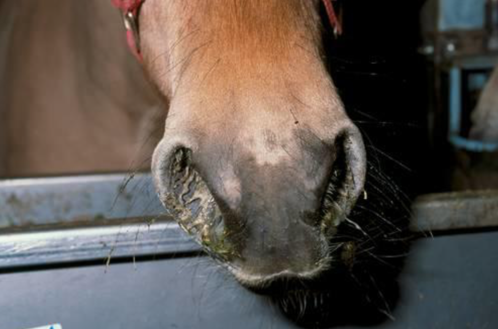
  • A clear or white nasal discharge is often seen with enlargement of the submandibular lymph nodes (under their chin/throat area).

Vaccinated horses

  • Horses that have been regularly vaccinated often show no clinical signs, but they may still shed enough virus to infect other horses. This is how the outbreak in Australia in 2007 started.
  • Horses that have been vaccinated but only have partial protection, e.g. because they haven’t been vaccinated frequently enough or because the vaccine type used was not updated, may show varied signs of mild non specific respiratory disease. This can include mild lethargy, nasal discharge and possibly a cough.

What to do if you think there is a possible infected horse?

Owners should seek veterinary advice if they suspect there is flu on the yard and carry out the following precautions:

  • Isolate the possibly infected horses. Infected horses will spread the virus in respiratory droplets, through coughing, and can spread the virus for up to 10 days. They must be isolated until the vet advises they are no longer infected.
  • All horse movements on and off the yard should be stopped.
  • Monitor all horses on the yard for clinical signs and record their rectal temperature daily, it should be less than 38.0˚C. A rise in temperature can be an early sign of an infection.
  • Booster vaccinating all in-contact horses, even if they are not yet due their annual booster, has been shown to provide horses with even more protection against flu.

How to prevent it

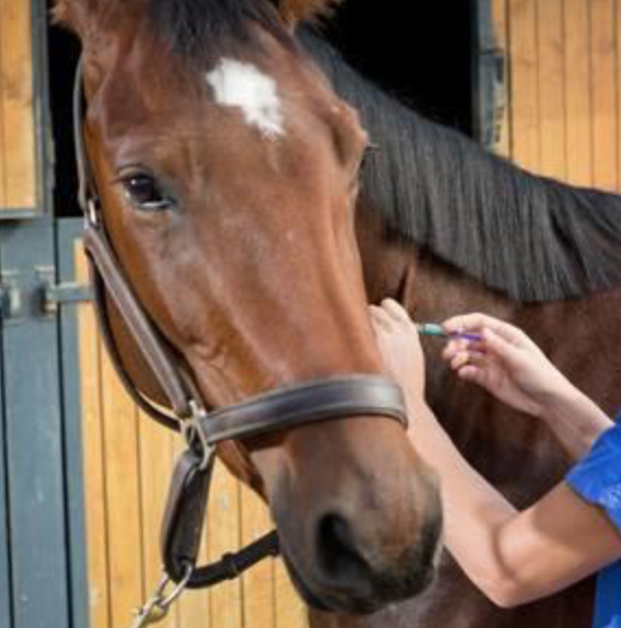
Horse Flu is endemic in the UK, which means that most horses will come into regular contact with the virus during their lifetime. The control of horse flu in the UK is based on limiting the signs of infection in horses that have been exposed to the virus, rather than trying to prevent exposure itself. This is primarily achieved through regular vaccination which is given either once or twice a year.
If you have any questions about your horse’s vaccinations, please call our Equine Team on 01772 861300

Free* ACTH tests are back for 2019

Testing for PPID in your horse or pony

A few years ago PPID or Equine Cushing’s Disease was considered a rare hormonal disease in horses. Now it is thought to affect over 20% of horses over the age of 15 and is a condition recognised almost daily in equine veterinary practice.

Past ‘Talk about Laminitis’ test results show that you should look out for any of these clinical signs in your horse:

  • Laminitis
  • Abnormal or delayed moulting
  • Muscle wastage
  • Periorbital fat (fat round the eyes)
  • Increased drinking and urinating

In 2017, thousands of laminitic patients benefitted from PPID being ruled in or out of their condition during the Talk About Laminitis campaign, as a result, the Talk About Laminitis campaign will continue offering FREE* ACTH lab testing during 2019

You can claim your complimentary testing voucher code by visiting www.careaboutcushings.co.uk

If your horse is already on Prascend, you may be eligible for a free monitoring test (lab fees only). You can generate your voucher code for this by visiting the link above.


Terms & Conditions:

  • This free ACTH test voucher code is for the laboratory fees for a blood ACTH test only.
  • Each voucher is only redeemable against an ACTH blood test where the patient has not been previously diagnosed with PPID
  • Full T&Cs can be found below at www.careaboutcushings.co.uk
  • Oakhill blood sampling fee, visit fee and postage still apply.

Winter Series: Mud Fever

Mud fever, also known as greasy heel syndrome but correctly termed pastern dermatitis, is an infection of the skin usually of the pastern.

The bacteria causing the infection can be from the environment or living on the skin itself and will gain entry due to an injury to the skin surface. It more commonly affects white socked legs, as the skin’s immune function of these areas is reduced. In minor cases it may present as just a few scabs, however, it can spread above the fetlock, occur concurrently with cellulitis, or leg filling, and be openly bleeding.

Although it can have a typical appearance sometimes a vet visit is needed to rule out other conditions and to identify predisposing factors.

The skins integrity is weakened by continuous wetting of the skin and rubbing from mud meaning it is a problem commonly seen in Winter. However, it may not just be wet and muddy conditions that are causing the problem……..

[checklist icon=”fa-arrow-circle-right” iconcolor=”#39aa87″ circle=”yes”]

  • Lymphoedema: this is typical of cob type or draught breeds. Lymph fluid circulates around the body carrying nutrients in lymph vessels. The lymph vessels in these types seem to be less effective in circulating blood in the lower limbs which leads to accumulation of fluid and skin thickening.
  • Leucocytoclastic vasculitis: this is an immune mediated condition whereby blood vessels become damaged. Personal experience shows it occurs more commonly on the outside of white pasterns.
  • Photosensitisation: this is caused by either liver disease or allergic reaction meaning the horse doesn’t deal with chemicals that react with light meaning that when the horse is exposed to UV radiation (sunlight) it becomes sunburnt.
  • Feather mites/ Chorioptic mange: Chorioptes mites causes itchiness of the lower limbs. This can lead to self trauma and wounds allowing skin infection.

[/checklist]

To rule out these conditions it may be necessary to take blood samples or biopsies to determine if one of these are involved.

In order to treat Mud Fever you may need to treat the underlying cause. Sometimes a thorough antibacterial cleaning and removal of scabs is needed, under sedation, so that topical antibacterial, steroidal and protectant creams can be applied directly onto the damaged skin. In some cases when associated with cellulitis or more widespread infection of the leg systemic antibiotics may be needed.

The deliberation is always; do you wash the mud off your horses legs or do you brush it off when dry ?? In our opinion neither is superior. If you are to wash your horses legs they need to be thoroughly dried and if there is already some mud fever present avoid wetting the leg further.

If you are concerned about Mud Fever in your horse, please call us on 01772 861300

Beef & Youngstock – Housing Considerations

We have experienced a drier summer than we are used to and this has posed different challenges. Worm burdens have been greatly reduced on pasture over the summer however if we get a heavy rainfall following this dry period there is likely to be mass emergence of worms onto pasture which may have severe consequences for our livestock if left untreated.

Exposure to lungworm may also have been reduced over the summer and so we would advise watching out for or listening to cattle to see if they are coughing over the next few months and treating if necessary. Lungworm can cause significant respiratory disease and death in cattle and should not be overlooked in any grazed stock. Where cattle have been grazed for extended periods the use of long acting anthelmintic bolus activity may have worn off and further treatment may be necessary.

Testing for Lungworm larvae can be done on a faecal sample however is slightly different to a normal worm egg count. If you would like to check grazed stock for Lungworm please submit a faecal sample stating that you require Lungworm testing.

Bringing in cattle from markets, other farms or common grazing can introduce new diseases to your own farm, including parasites such as liver fluke. Liver fluke is a common parasite that infects cattle, sheep and other domestic animals as well as wildlife such as deer and rabbits. Following several wet years monitoring, testing and treating for liver fluke early is also recommended during this year.

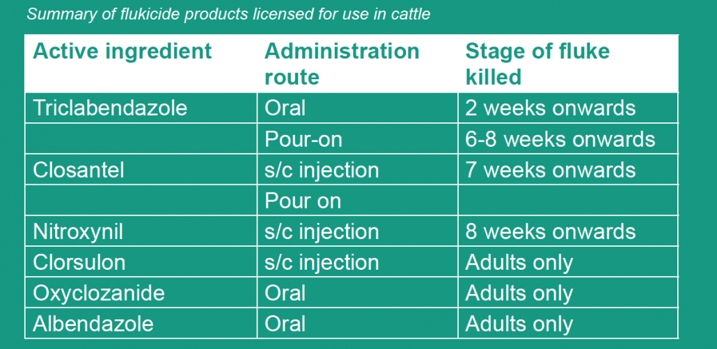
Resistance of liver fluke to treatments, in particular products containing triclabendazole (TCBZ), appears to be an increasing problem and one that needs managing on all livestock farms. It is important that care is taken to reduce selection pressure for resistance whenever possible by only using triclabendazole-containing products when no other options are suitable. Other medicines which contain the active ingredients, closantel, clorsulon, nitroxynil, oxyclozanide and albendazole are effective against adult flukes and some (nitroxynil and closantel) are effective against late immature stages between seven and eight weeks old.

If treating now or immediately on housing we would recommend using a Triclabendazole product but if cattle are housed for 7 weeks or more, one of the other products is often more suitable. Please speak to us about testing or when to treat.

Embryo Transfer

Embryo transfer- repeat breeder embryos (‘cow stoppers’)

What is a Problem Breeder Cow?
Cows that have had three or more services at regular (21-24 day) intervals and that are still not in calf.

What treatments can we use to treat problem breeders?
Problem Breeder Embryos – implanting a beef cross embryo 7 days after service in problem repeat breeder cows improved pregnancy rates by over 20% according to a recent UK study.

How does implanting an embryo help?
By implanting a second embryo into the cows’ uterus the strength of the hormonal signals within the cow are doubled and so the likelihood of the cow reabsorbing the embryo(s) is reduced.

What cows would make good Embryo Transfer candidates?

  • Cows should be cycling normally, showing regular heats every 21 – 24 days.
  • Cows should be vet-checked to make sure there is no uterine damage and no ovarian problems such as cystic ovaries.
  • Cows should be clean with no uterine discharge.
  • Ideally cows should be in reasonable body condition and good health. Chronically lame or high SCC cows are not ideal candidates!

What is the procedure?

  • Cow AI’d as normal. Arrange vet visit for 7 days later.
  • Cow examined prior to implanting the embryo.
  • We administer an epidural as this facilitates the procedure and should improve success rates.
  • Embryo implanted and cow scanned 5 weeks later to check if successful.

Maximising success rates in embryo transfer 
These are some suggestions when dealing with embryo recipients.

  • Maiden heifers make the best recipients: they have higher conception rates.
  • Cows can be used providing they have a good body condition score, and they are clean and cycling.
  • Embryos can be put in to heifers that were bulling 6, 7 or 8 days previously. The highest conception rates are likely to be achieved 7 days after a heat.
  • A good reference heat is vital to the success of the transfer.
  • Reference heats can be a natural observed heat or synchronised induced heats.
  • Included heats (i.e. Following a CIDR and prostaglandin programme) work equally well providing good heat detection is undertaken.
  • A wrong or inaccurate heat observation will mean no pregnancy as these recipients are not synchronised with the donor cow.
  • Avoid feeding changes to the recipient group for four to six weeks either side of transfers. Try to keep the diet consistent.
  • Knowing the trace element history of your farm is important.
  • Grass variation and grass silage variation can affect conception rates.